• The Indian Context: Intracranial Hemorrhage Care in Pune and Beyond

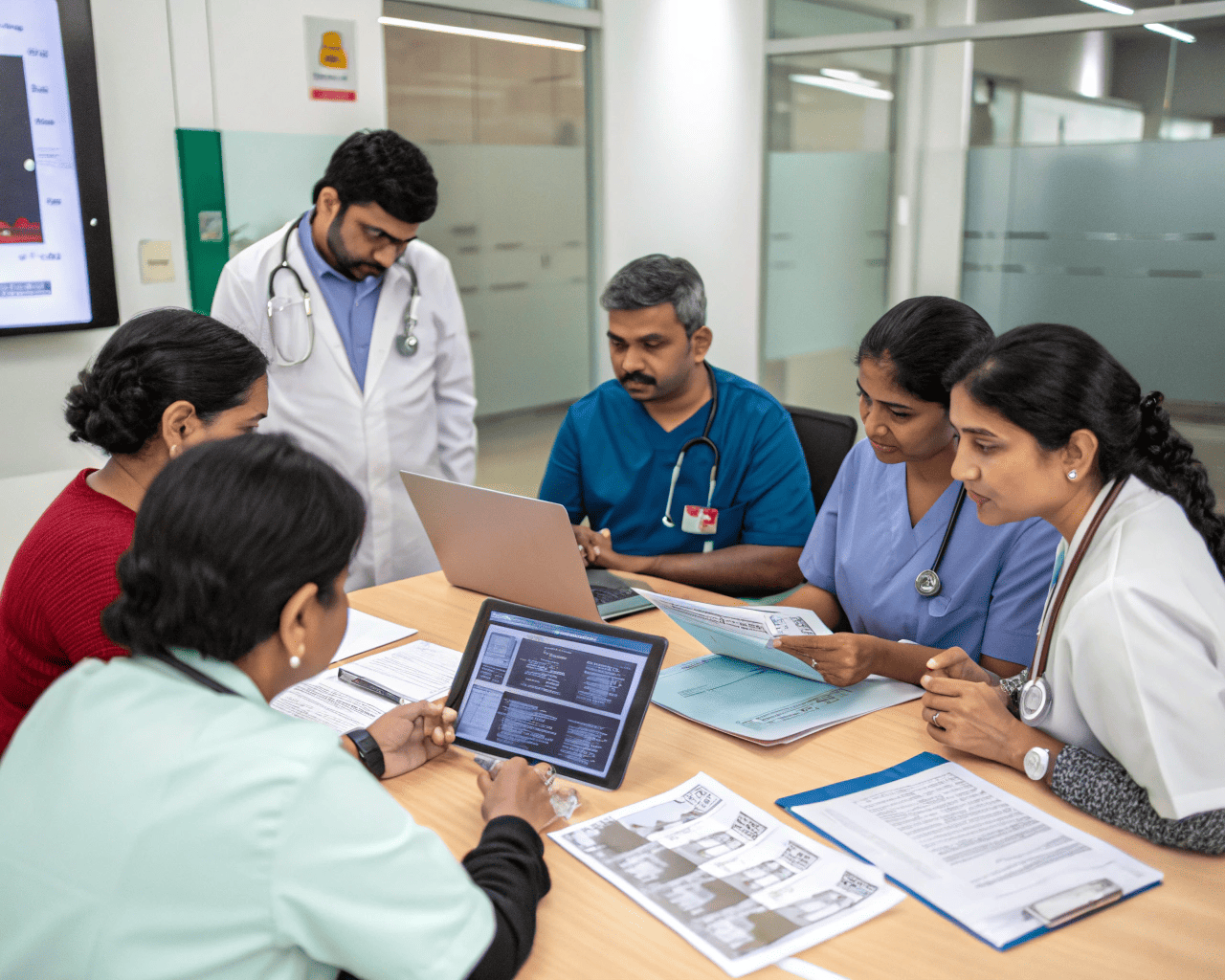
    In India, Intracranial Hemorrhage (ICH) presents a significant public health challenge, driven by factors such as a high prevalence of uncontrolled hypertension, increasing rates of road traffic accidents (RTAs), and an aging population.
    https://www.marketresearchfuture.com/reports/intracranial-hemorrhage-diagnosis-and-treatment-market-3687

    While major metropolitan cities like Pune boast advanced neurological care, disparities in access and awareness remain. Understanding the unique aspects of ICH diagnosis and treatment in the Indian context is crucial for improving outcomes.

    Prevalence and Causes in India:

    Hypertension: Uncontrolled hypertension is the leading cause of spontaneous (non-traumatic) ICH in India, mirroring global trends. Late diagnosis of hypertension and poor adherence to medication contribute to this burden.

    Trauma: India has one of the highest rates of road traffic accidents globally. Traumatic brain injuries (TBIs), a major cause of epidural and subdural hematomas, are a common presentation in emergency departments, especially in urban centers like Pune, which have high vehicular density.

    Rural-Urban Divide: While awareness and access to healthcare infrastructure are improving in urban areas, rural populations often face significant challenges in reaching specialized care quickly, leading to delays in diagnosis and treatment.

    Diagnostic Landscape in Pune:

    Pune, being a major economic and educational hub in Maharashtra, has a well-developed healthcare infrastructure, particularly in neurosciences.

    Accessibility of CT Scans: Most multi-specialty hospitals and large diagnostic centers in Pune are equipped with modern CT scanners, ensuring rapid diagnosis of acute ICH. This accessibility is vital for emergency management.

    Neurology and Neurosurgery Centers: Pune boasts several tertiary care hospitals with dedicated neurology and neurosurgery departments, offering advanced diagnostic capabilities (e.g., MRI, CTA, DSA) and experienced specialists. Hospitals such as Sahyadri Hospital, Apollo Hospitals, Ruby Hall Clinic, Jehangir Hospital, and Manipal Hospital are recognized for their neurological services and handle a high volume of ICH cases.

    Specialized Expertise: Neurosurgeons and neurologists in Pune are adept at diagnosing and managing all types of ICH, including complex cases requiring advanced surgical techniques or endovascular interventions for aneurysms and AVMs.

    Treatment Approaches in India:

    Emergency Response: The emphasis in urban centers like Pune is on rapid transport to an equipped hospital, immediate resuscitation, and urgent imaging. This "golden hour" approach is critical for minimizing brain damage.

    Medical Management: Management of blood pressure, intracranial pressure (ICP), and seizure prophylaxis follows international guidelines. However, affordability of high-cost medications or advanced monitoring devices can sometimes be a concern for patients from lower socioeconomic strata.

    Surgical Intervention: Access to neurosurgical expertise and operating facilities for craniotomy, hematoma evacuation, and aneurysm clipping/coiling is readily available in Pune's major hospitals. The decision for surgery is made based on standard criteria (hematoma size, location, neurological status) adapted to the specific patient context.

    Rehabilitation: Post-acute rehabilitation is increasingly recognized as crucial. Pune has a growing number of rehabilitation centers offering physical, occupational, and speech therapy, though comprehensive, long-term rehabilitation remains a challenge for many patients due to cost and family support structures.

    Challenges in the Indian Context:

    Pre-hospital Delay: Delays in recognizing symptoms and reaching a medical facility, especially from rural or semi-urban areas to specialized centers in Pune, can significantly worsen outcomes.

    Financial Burden: The cost of advanced diagnostic tests, emergency surgery, prolonged ICU stays, and long-term rehabilitation can be substantial, often leading to catastrophic out-of-pocket expenses for families without adequate health insurance.

    Awareness: Lower public awareness about stroke symptoms and the importance of immediate medical attention (Act FAST principles) contributes to treatment delays.

    Resource Disparities: While Pune is well-equipped, smaller towns and rural areas across India often lack the necessary neurosurgical facilities, ICU beds, and trained personnel.

    Post-Discharge Care: Ensuring continued medication adherence, follow-up, and access to rehabilitation services after discharge remains a challenge for many, impacting long-term recovery.

    Despite these challenges, India, and particularly cities like Pune, are making significant strides in improving ICH care. Increased public awareness campaigns, government health schemes, and the continuous upgrading of medical infrastructure are essential steps to bridge the existing gaps and provide equitable access to life-saving treatment for intracranial hemorrhage across the nation.
    The Indian Context: Intracranial Hemorrhage Care in Pune and Beyond In India, Intracranial Hemorrhage (ICH) presents a significant public health challenge, driven by factors such as a high prevalence of uncontrolled hypertension, increasing rates of road traffic accidents (RTAs), and an aging population. https://www.marketresearchfuture.com/reports/intracranial-hemorrhage-diagnosis-and-treatment-market-3687 While major metropolitan cities like Pune boast advanced neurological care, disparities in access and awareness remain. Understanding the unique aspects of ICH diagnosis and treatment in the Indian context is crucial for improving outcomes. Prevalence and Causes in India: Hypertension: Uncontrolled hypertension is the leading cause of spontaneous (non-traumatic) ICH in India, mirroring global trends. Late diagnosis of hypertension and poor adherence to medication contribute to this burden. Trauma: India has one of the highest rates of road traffic accidents globally. Traumatic brain injuries (TBIs), a major cause of epidural and subdural hematomas, are a common presentation in emergency departments, especially in urban centers like Pune, which have high vehicular density. Rural-Urban Divide: While awareness and access to healthcare infrastructure are improving in urban areas, rural populations often face significant challenges in reaching specialized care quickly, leading to delays in diagnosis and treatment. Diagnostic Landscape in Pune: Pune, being a major economic and educational hub in Maharashtra, has a well-developed healthcare infrastructure, particularly in neurosciences. Accessibility of CT Scans: Most multi-specialty hospitals and large diagnostic centers in Pune are equipped with modern CT scanners, ensuring rapid diagnosis of acute ICH. This accessibility is vital for emergency management. Neurology and Neurosurgery Centers: Pune boasts several tertiary care hospitals with dedicated neurology and neurosurgery departments, offering advanced diagnostic capabilities (e.g., MRI, CTA, DSA) and experienced specialists. Hospitals such as Sahyadri Hospital, Apollo Hospitals, Ruby Hall Clinic, Jehangir Hospital, and Manipal Hospital are recognized for their neurological services and handle a high volume of ICH cases. Specialized Expertise: Neurosurgeons and neurologists in Pune are adept at diagnosing and managing all types of ICH, including complex cases requiring advanced surgical techniques or endovascular interventions for aneurysms and AVMs. Treatment Approaches in India: Emergency Response: The emphasis in urban centers like Pune is on rapid transport to an equipped hospital, immediate resuscitation, and urgent imaging. This "golden hour" approach is critical for minimizing brain damage. Medical Management: Management of blood pressure, intracranial pressure (ICP), and seizure prophylaxis follows international guidelines. However, affordability of high-cost medications or advanced monitoring devices can sometimes be a concern for patients from lower socioeconomic strata. Surgical Intervention: Access to neurosurgical expertise and operating facilities for craniotomy, hematoma evacuation, and aneurysm clipping/coiling is readily available in Pune's major hospitals. The decision for surgery is made based on standard criteria (hematoma size, location, neurological status) adapted to the specific patient context. Rehabilitation: Post-acute rehabilitation is increasingly recognized as crucial. Pune has a growing number of rehabilitation centers offering physical, occupational, and speech therapy, though comprehensive, long-term rehabilitation remains a challenge for many patients due to cost and family support structures. Challenges in the Indian Context: Pre-hospital Delay: Delays in recognizing symptoms and reaching a medical facility, especially from rural or semi-urban areas to specialized centers in Pune, can significantly worsen outcomes. Financial Burden: The cost of advanced diagnostic tests, emergency surgery, prolonged ICU stays, and long-term rehabilitation can be substantial, often leading to catastrophic out-of-pocket expenses for families without adequate health insurance. Awareness: Lower public awareness about stroke symptoms and the importance of immediate medical attention (Act FAST principles) contributes to treatment delays. Resource Disparities: While Pune is well-equipped, smaller towns and rural areas across India often lack the necessary neurosurgical facilities, ICU beds, and trained personnel. Post-Discharge Care: Ensuring continued medication adherence, follow-up, and access to rehabilitation services after discharge remains a challenge for many, impacting long-term recovery. Despite these challenges, India, and particularly cities like Pune, are making significant strides in improving ICH care. Increased public awareness campaigns, government health schemes, and the continuous upgrading of medical infrastructure are essential steps to bridge the existing gaps and provide equitable access to life-saving treatment for intracranial hemorrhage across the nation.
    WWW.MARKETRESEARCHFUTURE.COM
    Intracranial Hemorrhage Diagnosis & Treatment Market by Type, Growth and Forecast – 2032
    Intracranial Hemorrhage Diagnosis & Treatment Market can advance at 7.2% CAGR by 2032, Global Intracranial Hemorrhage Diagnosis & Treatment Market categorizes the Worldwide Market by Type and Region | Intracranial Hemorrhage Diagnosis & Treatment Industry
    0 Comments 0 Shares
  • The Rise of Wearables: Monitoring Pediatric Neurology at Home

    In pediatric neurology, where conditions like epilepsy, cerebral palsy, and developmental disorders often require continuous monitoring, the traditional reliance on hospital visits and intermittent assessments can be burdensome and provide an incomplete picture.
    https://www.marketresearchfuture.com/reports/global-pediatric-neurology-devices-market-565

    The emergence of wearable devices is revolutionizing this landscape, allowing for real-time, continuous, and objective data collection in a child's natural environment—their home.

    These pediatric neurology devices are not only enhancing diagnostic capabilities but also empowering families and improving the management of chronic neurological conditions.

    Why Wearables are Game-Changers in Pediatric Neurology:

    Continuous, Real-World Data: Unlike clinical visits that capture a snapshot in time, wearables collect data 24/7. This continuous stream provides a more comprehensive understanding of symptom frequency, severity, and patterns, which can fluctuate throughout the day or night. For example, a child's seizure burden might be significantly underestimated if only relying on caregiver recall during clinic visits.

    Objective Measurement: Wearables often utilize sensors (accelerometers, gyroscopes, heart rate monitors, electrodermal activity sensors) to objectively quantify movements, physiological responses, and sleep. This reduces the subjectivity inherent in parent diaries or clinical rating scales.

    Early Detection and Warning: For conditions like epilepsy, some wearables can detect subtle changes indicative of an impending seizure or even alert caregivers during a seizure, potentially improving safety and enabling timely intervention.

    Remote Monitoring and Telemedicine: Wearable data can be transmitted wirelessly to healthcare providers, facilitating remote monitoring. This is particularly beneficial for families in remote areas or those with limited access to specialized pediatric neurologists, reducing the need for frequent, arduous hospital visits. In a country like India, with vast geographical disparities in healthcare access, this aspect holds immense potential.

    Personalized Treatment Adjustments: The rich data from wearables can help clinicians fine-tune medication dosages, adjust therapy plans, and assess the effectiveness of interventions more precisely, leading to more personalized and effective care.

    Empowering Families: Wearables can empower parents by providing insights into their child's condition, potentially reducing anxiety and increasing their sense of control. They can also serve as a tool for communication between parents and healthcare providers.
    Key Applications of Wearable Devices in Pediatric Neurology:

    Epilepsy Monitoring:
    Seizure Detection: Wearable EEG patches, smartwatches, or devices worn on the wrist or ankle can detect convulsive seizures by analyzing movement patterns (accelerometry) or changes in skin conductance (electrodermal activity, EDA), heart rate, or brainwaves.

    Devices like Empatica Embrace2 are examples that have received regulatory clearance for seizure detection in some regions.
    Sleep Monitoring: Tracking sleep patterns can provide valuable information, as sleep disturbances are common in children with epilepsy and can influence seizure frequency.

    Cerebral Palsy and Movement Disorders:
    Motor Function Assessment: Accelerometers and gyroscopes in wearables can objectively measure gait, balance, tremor, and limb usage, helping therapists track progress, tailor rehabilitation exercises, and assess functional improvements over time.
    Activity Monitoring: Quantifying daily activity levels and movement patterns provides insights into a child's participation in daily life.

    Autism Spectrum Disorder (ASD):
    Physiological Monitoring: Wearables can track physiological arousal (e.g., heart rate, EDA) that may correlate with anxiety or stress, providing objective data to understand triggers and manage challenging behaviors.

    Sleep Disorders: Wearable sleep trackers can monitor sleep cycles, quality, and disturbances in children with neurological conditions, guiding appropriate interventions.

    Challenges and the Indian Outlook:

    Despite their promise, challenges remain. These include ensuring accuracy and reliability, user compliance (especially for young children), data security and privacy, device cost, and regulatory hurdles for medical-grade wearables. In India, affordability and the digital divide (access to smartphones/internet for data transmission) are significant considerations.

    However, the rapidly expanding smartphone penetration and increasing tech-savviness are paving the way for wider adoption. Local innovations focused on cost-effectiveness and culturally appropriate designs will be key to unlocking the full potential of wearables in pediatric neurology across India.

    Wearable devices are no longer just gadgets; they are becoming indispensable tools in pediatric neurology, offering a continuous, objective window into a child's neurological health and transforming how chronic conditions are monitored and managed at home.
    The Rise of Wearables: Monitoring Pediatric Neurology at Home In pediatric neurology, where conditions like epilepsy, cerebral palsy, and developmental disorders often require continuous monitoring, the traditional reliance on hospital visits and intermittent assessments can be burdensome and provide an incomplete picture. https://www.marketresearchfuture.com/reports/global-pediatric-neurology-devices-market-565 The emergence of wearable devices is revolutionizing this landscape, allowing for real-time, continuous, and objective data collection in a child's natural environment—their home. These pediatric neurology devices are not only enhancing diagnostic capabilities but also empowering families and improving the management of chronic neurological conditions. Why Wearables are Game-Changers in Pediatric Neurology: Continuous, Real-World Data: Unlike clinical visits that capture a snapshot in time, wearables collect data 24/7. This continuous stream provides a more comprehensive understanding of symptom frequency, severity, and patterns, which can fluctuate throughout the day or night. For example, a child's seizure burden might be significantly underestimated if only relying on caregiver recall during clinic visits. Objective Measurement: Wearables often utilize sensors (accelerometers, gyroscopes, heart rate monitors, electrodermal activity sensors) to objectively quantify movements, physiological responses, and sleep. This reduces the subjectivity inherent in parent diaries or clinical rating scales. Early Detection and Warning: For conditions like epilepsy, some wearables can detect subtle changes indicative of an impending seizure or even alert caregivers during a seizure, potentially improving safety and enabling timely intervention. Remote Monitoring and Telemedicine: Wearable data can be transmitted wirelessly to healthcare providers, facilitating remote monitoring. This is particularly beneficial for families in remote areas or those with limited access to specialized pediatric neurologists, reducing the need for frequent, arduous hospital visits. In a country like India, with vast geographical disparities in healthcare access, this aspect holds immense potential. Personalized Treatment Adjustments: The rich data from wearables can help clinicians fine-tune medication dosages, adjust therapy plans, and assess the effectiveness of interventions more precisely, leading to more personalized and effective care. Empowering Families: Wearables can empower parents by providing insights into their child's condition, potentially reducing anxiety and increasing their sense of control. They can also serve as a tool for communication between parents and healthcare providers. Key Applications of Wearable Devices in Pediatric Neurology: Epilepsy Monitoring: Seizure Detection: Wearable EEG patches, smartwatches, or devices worn on the wrist or ankle can detect convulsive seizures by analyzing movement patterns (accelerometry) or changes in skin conductance (electrodermal activity, EDA), heart rate, or brainwaves. Devices like Empatica Embrace2 are examples that have received regulatory clearance for seizure detection in some regions. Sleep Monitoring: Tracking sleep patterns can provide valuable information, as sleep disturbances are common in children with epilepsy and can influence seizure frequency. Cerebral Palsy and Movement Disorders: Motor Function Assessment: Accelerometers and gyroscopes in wearables can objectively measure gait, balance, tremor, and limb usage, helping therapists track progress, tailor rehabilitation exercises, and assess functional improvements over time. Activity Monitoring: Quantifying daily activity levels and movement patterns provides insights into a child's participation in daily life. Autism Spectrum Disorder (ASD): Physiological Monitoring: Wearables can track physiological arousal (e.g., heart rate, EDA) that may correlate with anxiety or stress, providing objective data to understand triggers and manage challenging behaviors. Sleep Disorders: Wearable sleep trackers can monitor sleep cycles, quality, and disturbances in children with neurological conditions, guiding appropriate interventions. Challenges and the Indian Outlook: Despite their promise, challenges remain. These include ensuring accuracy and reliability, user compliance (especially for young children), data security and privacy, device cost, and regulatory hurdles for medical-grade wearables. In India, affordability and the digital divide (access to smartphones/internet for data transmission) are significant considerations. However, the rapidly expanding smartphone penetration and increasing tech-savviness are paving the way for wider adoption. Local innovations focused on cost-effectiveness and culturally appropriate designs will be key to unlocking the full potential of wearables in pediatric neurology across India. Wearable devices are no longer just gadgets; they are becoming indispensable tools in pediatric neurology, offering a continuous, objective window into a child's neurological health and transforming how chronic conditions are monitored and managed at home.
    WWW.MARKETRESEARCHFUTURE.COM
    Pediatric Neurology Devices Market Size, Share, Trends 2032 | MRFR
    Pediatric Neurology Devices Market to grow at a CAGR over 5.12% By 2032. Pediatric Neurology Devices Market by Type, Treatment, Age Group and End user | Pediatric Neurology Devices Industry | MRFR
    0 Comments 0 Shares
  • Living with LAM: Navigating Life with a Rare Lung Disease in India

    A diagnosis of Lymphangioleiomyomatosis (LAM) is life-altering. For women in India, grappling with a rare, chronic, and progressive lung disease brings a unique set of challenges beyond the clinical symptoms.
    https://www.marketresearchfuture.com/reports/lymphangioleiomyomatosis-market-21872

    However, with appropriate management, lifestyle adjustments, and strong support systems, individuals with LAM can lead fulfilling lives and effectively navigate their condition.
    https://www.marketresearchfuture.com/reports/lymphangioleiomyomatosis-market-21872

    Understanding the Impact on Daily Life:

    LAM primarily impacts the lungs, leading to:

    Breathlessness: This is the most common and often debilitating symptom. It can restrict physical activity, affecting daily chores, work, and social interactions.

    Fatigue: Chronic fatigue is prevalent and can significantly reduce energy levels.

    Recurrent Pneumothorax: The fear of a collapsed lung can create anxiety and impact travel plans or participation in certain activities.

    Medical Appointments and Medications: Regular doctor visits, diagnostic tests, and daily medication adherence become a routine part of life.

    Beyond the physical, LAM can also take a toll on mental and emotional well-being:

    Anxiety and Depression: The uncertainty of a progressive disease, the fear of exacerbations, and the impact on daily life can lead to significant psychological distress.

    Social Isolation: The limitations imposed by breathlessness or fear of complications can lead to reduced social engagement.

    Impact on Work and Family: LAM often strikes women in their reproductive and peak career years, affecting family planning, professional aspirations, and financial stability.
    Strategies for Managing Life with LAM in India:

    Adherence to Treatment and Monitoring:

    Medication Consistency: Taking sirolimus or other prescribed medications as directed is paramount. Understanding dosage, potential side effects, and the importance of regular blood level monitoring for sirolimus is crucial.

    Regular Follow-ups: Consistent appointments with a pulmonologist and other specialists are essential for tracking disease progression, adjusting treatment, and managing complications.

    Self-Monitoring: Patients should be educated on recognizing worsening symptoms and when to seek urgent medical attention.
    Pulmonary Rehabilitation:

    This is an invaluable component of care. Pulmonary rehab programs (exercise training, breathing techniques, and education) can significantly improve lung function, exercise capacity, and overall quality of life, even if lung function numbers don't drastically improve.

    Indian Context: While not widely available everywhere, seeking out centers in major cities that offer pulmonary rehabilitation can make a substantial difference. Home-based exercise programs with professional guidance can also be beneficial.

    Lifestyle Modifications:

    Avoid Smoking: Absolutely critical, as smoking can severely worsen lung damage.

    Healthy Diet: A balanced diet supports overall health and energy levels. For patients with chylous effusions, a low-fat diet may be recommended.

    Stay Active (within limits): Regular, moderate exercise, tailored to individual capacity, can help maintain muscle strength and cardiovascular health.

    Avoid High-Risk Activities: Activities that involve significant pressure changes (e.g., scuba diving) should be avoided due to the risk of pneumothorax.

    Mental and Emotional Well-being:

    Counselling and Support: Seeking professional counseling can help individuals cope with the emotional burden of a chronic illness.

    Patient Support Groups: Connecting with other LAM patients, either online or in-person, provides invaluable emotional support, shared experiences, and practical advice. The global LAM Foundation is an excellent resource, and efforts to build similar networks in India are vital.

    Stress Management: Techniques like yoga, meditation, or mindfulness can help manage stress and anxiety.

    Navigating the Indian Healthcare System:

    Seek Specialized Care: Identifying and connecting with pulmonologists and centers with experience in rare lung diseases is crucial.

    Financial Planning: Understanding potential costs and exploring available government schemes or patient assistance programs for rare diseases can help alleviate financial stress.

    Advocacy: Patients and their families may need to advocate for their needs, whether it's for access to specific tests, medications, or specialized consultations.

    Living with LAM in India requires resilience, informed decision-making, and proactive engagement with the healthcare system.

    While the journey can be challenging, the growing scientific understanding, available treatments, and power of community support offer hope and enable individuals with LAM to lead meaningful and productive lives.
    Living with LAM: Navigating Life with a Rare Lung Disease in India A diagnosis of Lymphangioleiomyomatosis (LAM) is life-altering. For women in India, grappling with a rare, chronic, and progressive lung disease brings a unique set of challenges beyond the clinical symptoms. https://www.marketresearchfuture.com/reports/lymphangioleiomyomatosis-market-21872 However, with appropriate management, lifestyle adjustments, and strong support systems, individuals with LAM can lead fulfilling lives and effectively navigate their condition. https://www.marketresearchfuture.com/reports/lymphangioleiomyomatosis-market-21872 Understanding the Impact on Daily Life: LAM primarily impacts the lungs, leading to: Breathlessness: This is the most common and often debilitating symptom. It can restrict physical activity, affecting daily chores, work, and social interactions. Fatigue: Chronic fatigue is prevalent and can significantly reduce energy levels. Recurrent Pneumothorax: The fear of a collapsed lung can create anxiety and impact travel plans or participation in certain activities. Medical Appointments and Medications: Regular doctor visits, diagnostic tests, and daily medication adherence become a routine part of life. Beyond the physical, LAM can also take a toll on mental and emotional well-being: Anxiety and Depression: The uncertainty of a progressive disease, the fear of exacerbations, and the impact on daily life can lead to significant psychological distress. Social Isolation: The limitations imposed by breathlessness or fear of complications can lead to reduced social engagement. Impact on Work and Family: LAM often strikes women in their reproductive and peak career years, affecting family planning, professional aspirations, and financial stability. Strategies for Managing Life with LAM in India: Adherence to Treatment and Monitoring: Medication Consistency: Taking sirolimus or other prescribed medications as directed is paramount. Understanding dosage, potential side effects, and the importance of regular blood level monitoring for sirolimus is crucial. Regular Follow-ups: Consistent appointments with a pulmonologist and other specialists are essential for tracking disease progression, adjusting treatment, and managing complications. Self-Monitoring: Patients should be educated on recognizing worsening symptoms and when to seek urgent medical attention. Pulmonary Rehabilitation: This is an invaluable component of care. Pulmonary rehab programs (exercise training, breathing techniques, and education) can significantly improve lung function, exercise capacity, and overall quality of life, even if lung function numbers don't drastically improve. Indian Context: While not widely available everywhere, seeking out centers in major cities that offer pulmonary rehabilitation can make a substantial difference. Home-based exercise programs with professional guidance can also be beneficial. Lifestyle Modifications: Avoid Smoking: Absolutely critical, as smoking can severely worsen lung damage. Healthy Diet: A balanced diet supports overall health and energy levels. For patients with chylous effusions, a low-fat diet may be recommended. Stay Active (within limits): Regular, moderate exercise, tailored to individual capacity, can help maintain muscle strength and cardiovascular health. Avoid High-Risk Activities: Activities that involve significant pressure changes (e.g., scuba diving) should be avoided due to the risk of pneumothorax. Mental and Emotional Well-being: Counselling and Support: Seeking professional counseling can help individuals cope with the emotional burden of a chronic illness. Patient Support Groups: Connecting with other LAM patients, either online or in-person, provides invaluable emotional support, shared experiences, and practical advice. The global LAM Foundation is an excellent resource, and efforts to build similar networks in India are vital. Stress Management: Techniques like yoga, meditation, or mindfulness can help manage stress and anxiety. Navigating the Indian Healthcare System: Seek Specialized Care: Identifying and connecting with pulmonologists and centers with experience in rare lung diseases is crucial. Financial Planning: Understanding potential costs and exploring available government schemes or patient assistance programs for rare diseases can help alleviate financial stress. Advocacy: Patients and their families may need to advocate for their needs, whether it's for access to specific tests, medications, or specialized consultations. Living with LAM in India requires resilience, informed decision-making, and proactive engagement with the healthcare system. While the journey can be challenging, the growing scientific understanding, available treatments, and power of community support offer hope and enable individuals with LAM to lead meaningful and productive lives.
    WWW.MARKETRESEARCHFUTURE.COM
    Lymphangioleiomyomatosis (LAM) Market Size, Share 2034 | MRFR
    Lymphangioleiomyomatosis (LAM) Market projected to grow at 5.51% CAGR, reaching USD 0.27 Billion by 2034. Top company industry analysis driving growth, trends, regions, opportunity, and global outlook 2025-2034.
    0 Comments 0 Shares
  • Enhancing Patient Mobility with MBT Device in Bhutan Hospitals

    Modern healthcare facilities are embracing mobility-focused rehabilitation tools, and the integration of the #MBT_device_in_Bhutan hospitals marks a significant step forward. Designed to aid in physical therapy and balance training, MBT (Mobile Balance Trainer) devices are improving recovery outcomes for patients with neurological and orthopedic conditions. With their user-friendly design and clinical accuracy, MBT devices are becoming an essential part of rehabilitation protocols across Bhutan’s medical centers.
    http://www.rshealthsupplies.com/mbt-therapy-machine-supplier
    Enhancing Patient Mobility with MBT Device in Bhutan Hospitals Modern healthcare facilities are embracing mobility-focused rehabilitation tools, and the integration of the #MBT_device_in_Bhutan hospitals marks a significant step forward. Designed to aid in physical therapy and balance training, MBT (Mobile Balance Trainer) devices are improving recovery outcomes for patients with neurological and orthopedic conditions. With their user-friendly design and clinical accuracy, MBT devices are becoming an essential part of rehabilitation protocols across Bhutan’s medical centers. http://www.rshealthsupplies.com/mbt-therapy-machine-supplier
    MBT Therapy Machine Supplier
    0 Comments 0 Shares
  • Beyond the Inhaler: Future Directions and Holistic Management of Airway Diseases

    Bronchodilators have undeniably transformed the lives of individuals with asthma and COPD, but the field of respiratory medicine continues to evolve. Future directions in bronchodilator therapy, combined with advancements in holistic patient care, aim to offer even greater precision, convenience, and comprehensive management of airway diseases.
    https://www.marketresearchfuture.com/reports/bronchodilators-market-8453

    Future Directions in Bronchodilator Therapy:

    Ultra-Long-Acting Bronchodilators:

    Innovation: While current LABAs and LAMAs offer 12-24 hour durations, research is exploring bronchodilators with even longer durations of action (e.g., once-weekly dosing).
    Impact: Further improve patient adherence by reducing dosing frequency, making it even easier for patients to stick to their treatment plans.

    Novel Bronchodilator Classes:

    Emerging Targets: Scientists are actively searching for new molecular targets and pathways involved in airway smooth muscle contraction and inflammation. This could lead to entirely new classes of bronchodilators with different mechanisms of action, potentially offering better efficacy or fewer side effects.
    Beyond Beta-2 and Muscarinic Receptors: Research is delving into compounds that modulate ion channels or other receptor systems in the airways.

    Smart Inhalers and Digital Health Integration:

    Connected Devices: "Smart inhalers" are already available, equipped with sensors that track usage patterns, remind patients to take their medication, and even transmit data to smartphones or healthcare providers.

    Personalized Insights: Future smart inhalers will offer more sophisticated insights, linking inhaler use to environmental triggers, symptom patterns, and even predicting exacerbations based on collected data.

    Telemedicine: Seamless integration with telehealth platforms will enable remote monitoring, virtual consultations, and personalized coaching, empowering patients in their self-management.

    Tailored Combination Therapies:

    Personalized Phenotyping/Endotyping: As our understanding of asthma and COPD subgroups (phenotypes/endotypes) grows, future therapies will be even more precisely matched to individual patients.

    This means selecting specific bronchodilator combinations (or combination with other drug classes like biologics) based on a patient's unique biological characteristics and inflammatory pathways.

    Quadruple Therapy: For very severe and difficult-to-treat cases, the concept of "quadruple therapy" (e.g., ICS + LABA + LAMA + another agent like a PDE4 inhibitor) is being explored for maximal benefit.
    Holistic Management Beyond Medication:

    While bronchodilators are vital, effective management of asthma and COPD requires a holistic approach that extends beyond pharmacological interventions:

    Pulmonary Rehabilitation: A comprehensive program of exercise training, education, and nutritional counseling, particularly crucial for COPD, significantly improves exercise capacity, reduces symptoms, and enhances quality of life.

    Smoking Cessation: For COPD, quitting smoking is the single most important intervention to slow disease progression.

    Trigger Avoidance: Identifying and avoiding environmental triggers (allergens, pollutants, irritants) for asthma, and exposure to respiratory irritants for COPD, is critical.

    Vaccinations: Annual flu shots and pneumonia vaccinations are essential to prevent exacerbations in both conditions.

    Patient Education and Self-Management: Empowering patients with knowledge about their condition, medication adherence, inhaler technique, and action plans for worsening symptoms is fundamental.

    Nutritional Support: Addressing nutritional deficiencies and promoting a healthy diet can support overall lung health.

    Psychological Support: Chronic respiratory conditions can take a toll on mental health. Addressing anxiety and depression is an important part of comprehensive care.

    The future of bronchodilator therapy, combined with these holistic strategies, aims to provide increasingly precise, convenient, and patient-centered care, enabling individuals with airway diseases to breathe easier and live fuller lives.
    Beyond the Inhaler: Future Directions and Holistic Management of Airway Diseases Bronchodilators have undeniably transformed the lives of individuals with asthma and COPD, but the field of respiratory medicine continues to evolve. Future directions in bronchodilator therapy, combined with advancements in holistic patient care, aim to offer even greater precision, convenience, and comprehensive management of airway diseases. https://www.marketresearchfuture.com/reports/bronchodilators-market-8453 Future Directions in Bronchodilator Therapy: Ultra-Long-Acting Bronchodilators: Innovation: While current LABAs and LAMAs offer 12-24 hour durations, research is exploring bronchodilators with even longer durations of action (e.g., once-weekly dosing). Impact: Further improve patient adherence by reducing dosing frequency, making it even easier for patients to stick to their treatment plans. Novel Bronchodilator Classes: Emerging Targets: Scientists are actively searching for new molecular targets and pathways involved in airway smooth muscle contraction and inflammation. This could lead to entirely new classes of bronchodilators with different mechanisms of action, potentially offering better efficacy or fewer side effects. Beyond Beta-2 and Muscarinic Receptors: Research is delving into compounds that modulate ion channels or other receptor systems in the airways. Smart Inhalers and Digital Health Integration: Connected Devices: "Smart inhalers" are already available, equipped with sensors that track usage patterns, remind patients to take their medication, and even transmit data to smartphones or healthcare providers. Personalized Insights: Future smart inhalers will offer more sophisticated insights, linking inhaler use to environmental triggers, symptom patterns, and even predicting exacerbations based on collected data. Telemedicine: Seamless integration with telehealth platforms will enable remote monitoring, virtual consultations, and personalized coaching, empowering patients in their self-management. Tailored Combination Therapies: Personalized Phenotyping/Endotyping: As our understanding of asthma and COPD subgroups (phenotypes/endotypes) grows, future therapies will be even more precisely matched to individual patients. This means selecting specific bronchodilator combinations (or combination with other drug classes like biologics) based on a patient's unique biological characteristics and inflammatory pathways. Quadruple Therapy: For very severe and difficult-to-treat cases, the concept of "quadruple therapy" (e.g., ICS + LABA + LAMA + another agent like a PDE4 inhibitor) is being explored for maximal benefit. Holistic Management Beyond Medication: While bronchodilators are vital, effective management of asthma and COPD requires a holistic approach that extends beyond pharmacological interventions: Pulmonary Rehabilitation: A comprehensive program of exercise training, education, and nutritional counseling, particularly crucial for COPD, significantly improves exercise capacity, reduces symptoms, and enhances quality of life. Smoking Cessation: For COPD, quitting smoking is the single most important intervention to slow disease progression. Trigger Avoidance: Identifying and avoiding environmental triggers (allergens, pollutants, irritants) for asthma, and exposure to respiratory irritants for COPD, is critical. Vaccinations: Annual flu shots and pneumonia vaccinations are essential to prevent exacerbations in both conditions. Patient Education and Self-Management: Empowering patients with knowledge about their condition, medication adherence, inhaler technique, and action plans for worsening symptoms is fundamental. Nutritional Support: Addressing nutritional deficiencies and promoting a healthy diet can support overall lung health. Psychological Support: Chronic respiratory conditions can take a toll on mental health. Addressing anxiety and depression is an important part of comprehensive care. The future of bronchodilator therapy, combined with these holistic strategies, aims to provide increasingly precise, convenient, and patient-centered care, enabling individuals with airway diseases to breathe easier and live fuller lives.
    WWW.MARKETRESEARCHFUTURE.COM
    Bronchodilators Market Size, Trends | Industry Analysis, 2032
    Bronchodilators Market to touch USD 53.44 Billion at 5.78% CAGR During the forecast period, Bronchodilators Industry Analysis by Drug Type, Disease, Mode of Action and Route of Administration
    0 Comments 0 Shares
  • Niramaya Oncology is widely regarded as the best oncology hospital in Gujarat, offering advanced cancer treatment with personalized care and cutting-edge technology. Our team of expert oncologists, surgeons, and support staff is dedicated to treating a wide range of cancers, including breast, lung, head and neck, and gastrointestinal cancers. We provide a full spectrum of services, from early diagnosis to post-treatment rehabilitation. With a patient-first approach and world-class infrastructure, we ensure holistic care and better outcomes. Choose Niramaya Oncology—the best oncology hospital in Gujarat—for compassionate treatment, modern therapies, and unwavering support through every stage of your cancer journey.

    Visit for more info:- https://niramayaoncology.com/
    Niramaya Oncology is widely regarded as the best oncology hospital in Gujarat, offering advanced cancer treatment with personalized care and cutting-edge technology. Our team of expert oncologists, surgeons, and support staff is dedicated to treating a wide range of cancers, including breast, lung, head and neck, and gastrointestinal cancers. We provide a full spectrum of services, from early diagnosis to post-treatment rehabilitation. With a patient-first approach and world-class infrastructure, we ensure holistic care and better outcomes. Choose Niramaya Oncology—the best oncology hospital in Gujarat—for compassionate treatment, modern therapies, and unwavering support through every stage of your cancer journey. Visit for more info:- https://niramayaoncology.com/
    0 Comments 0 Shares
  • The Road Ahead: Prognosis and Long-Term Outlook for Guillain-Barré Syndrome

    The diagnosis of Guillain-Barré Syndrome can be daunting, bringing with it fears of long-term disability or even death. However, it's important to understand that with prompt diagnosis and appropriate medical intervention, the prognosis for most individuals with GBS is generally positive, with the majority experiencing significant recovery.
    https://www.marketresearchfuture.com/reports/guillain-barre-syndrome-market-5741

    Recovery Timeline:
    Recovery from GBS is a gradual process, and the timeline varies widely among individuals.

    Initial Improvement: Most patients begin to show improvement within weeks or months after the peak of their weakness.
    Full Recovery: While some individuals recover completely within a few weeks to several months, others may take a year or longer for full recovery. Ongoing improvement can even be observed for up to two years or more after the initial attack.
    Factors Influencing Prognosis:
    Several factors can influence the overall prognosis and the extent of recovery:

    Severity of Initial Symptoms: Those with very severe weakness, especially requiring mechanical ventilation, may have a longer and more challenging recovery.
    Rapidity of Progression: Very rapid progression of symptoms can sometimes indicate a more severe course.
    Age: Older adults may tend to have a slower or less complete recovery compared to younger individuals.
    Type of GBS: While AIDP (demyelinating form) generally has a better prognosis, axonal forms (AMAN, AMSAN) can sometimes lead to more severe and prolonged weakness due to direct damage to the nerve fibers.
    Time to Treatment: Early administration of IVIG or plasmapheresis is crucial and significantly improves outcomes.
    Long-Term Effects and Complications:
    While most people recover well, approximately 15-30% of individuals may experience some long-term residual effects:

    Persistent Weakness or Numbness: This is the most common long-term issue, often affecting the feet and lower legs. It can range from mild tingling or numbness to more noticeable weakness that may require ongoing support (e.g., ankle braces, walking aids).

    Fatigue: Chronic fatigue is a significant and often underestimated long-term symptom, impacting quality of life for many survivors.
    Chronic Pain: Neuropathic pain, a type of nerve pain, can persist for some individuals.
    Balance and Coordination Issues: Due to lingering sensory and motor deficits.
    Autonomic Dysfunction: Less common, but some individuals may have lingering issues with blood pressure regulation, heart rate, or bowel/bladder function.
    Relapse: While rare, a small percentage (about 2-5%) of GBS patients may experience a relapse of symptoms, sometimes years after the initial recovery.
    Mortality Rate:
    GBS is rarely fatal, with a mortality rate estimated to be between 3% and 10%. Deaths, when they occur, are typically due to severe complications such as respiratory failure, severe autonomic dysfunction leading to cardiac arrest, or complications from prolonged immobility like pneumonia or blood clots. Prompt and intensive supportive care in an ICU plays a crucial role in reducing this mortality.

    Life Expectancy:
    For the vast majority of individuals who survive the acute phase and receive appropriate treatment, GBS does not significantly impact their overall life expectancy. With proper medical care and rehabilitation, many individuals can return to a fulfilling life, often with minimal or manageable long-term challenges.

    Ongoing Management and Support:
    For those with lingering symptoms, ongoing rehabilitation (physical, occupational, speech therapy) is vital to maximize recovery and adapt to any residual limitations. Managing fatigue and chronic pain often requires a multidisciplinary approach.

    Psychological support and counseling are also essential, as the sudden and debilitating nature of GBS can have a profound emotional impact on patients and their families. Support groups can provide invaluable shared experiences and coping strategies.

    In conclusion, while GBS presents a serious medical challenge, significant advancements in treatment and supportive care mean that most patients can anticipate a positive outcome, often regaining much of their previous function and quality of life.
    The Road Ahead: Prognosis and Long-Term Outlook for Guillain-Barré Syndrome The diagnosis of Guillain-Barré Syndrome can be daunting, bringing with it fears of long-term disability or even death. However, it's important to understand that with prompt diagnosis and appropriate medical intervention, the prognosis for most individuals with GBS is generally positive, with the majority experiencing significant recovery. https://www.marketresearchfuture.com/reports/guillain-barre-syndrome-market-5741 Recovery Timeline: Recovery from GBS is a gradual process, and the timeline varies widely among individuals. Initial Improvement: Most patients begin to show improvement within weeks or months after the peak of their weakness. Full Recovery: While some individuals recover completely within a few weeks to several months, others may take a year or longer for full recovery. Ongoing improvement can even be observed for up to two years or more after the initial attack. Factors Influencing Prognosis: Several factors can influence the overall prognosis and the extent of recovery: Severity of Initial Symptoms: Those with very severe weakness, especially requiring mechanical ventilation, may have a longer and more challenging recovery. Rapidity of Progression: Very rapid progression of symptoms can sometimes indicate a more severe course. Age: Older adults may tend to have a slower or less complete recovery compared to younger individuals. Type of GBS: While AIDP (demyelinating form) generally has a better prognosis, axonal forms (AMAN, AMSAN) can sometimes lead to more severe and prolonged weakness due to direct damage to the nerve fibers. Time to Treatment: Early administration of IVIG or plasmapheresis is crucial and significantly improves outcomes. Long-Term Effects and Complications: While most people recover well, approximately 15-30% of individuals may experience some long-term residual effects: Persistent Weakness or Numbness: This is the most common long-term issue, often affecting the feet and lower legs. It can range from mild tingling or numbness to more noticeable weakness that may require ongoing support (e.g., ankle braces, walking aids). Fatigue: Chronic fatigue is a significant and often underestimated long-term symptom, impacting quality of life for many survivors. Chronic Pain: Neuropathic pain, a type of nerve pain, can persist for some individuals. Balance and Coordination Issues: Due to lingering sensory and motor deficits. Autonomic Dysfunction: Less common, but some individuals may have lingering issues with blood pressure regulation, heart rate, or bowel/bladder function. Relapse: While rare, a small percentage (about 2-5%) of GBS patients may experience a relapse of symptoms, sometimes years after the initial recovery. Mortality Rate: GBS is rarely fatal, with a mortality rate estimated to be between 3% and 10%. Deaths, when they occur, are typically due to severe complications such as respiratory failure, severe autonomic dysfunction leading to cardiac arrest, or complications from prolonged immobility like pneumonia or blood clots. Prompt and intensive supportive care in an ICU plays a crucial role in reducing this mortality. Life Expectancy: For the vast majority of individuals who survive the acute phase and receive appropriate treatment, GBS does not significantly impact their overall life expectancy. With proper medical care and rehabilitation, many individuals can return to a fulfilling life, often with minimal or manageable long-term challenges. Ongoing Management and Support: For those with lingering symptoms, ongoing rehabilitation (physical, occupational, speech therapy) is vital to maximize recovery and adapt to any residual limitations. Managing fatigue and chronic pain often requires a multidisciplinary approach. Psychological support and counseling are also essential, as the sudden and debilitating nature of GBS can have a profound emotional impact on patients and their families. Support groups can provide invaluable shared experiences and coping strategies. In conclusion, while GBS presents a serious medical challenge, significant advancements in treatment and supportive care mean that most patients can anticipate a positive outcome, often regaining much of their previous function and quality of life.
    WWW.MARKETRESEARCHFUTURE.COM
    Guillain-Barre Syndrome Market Size, Growth, Analysis 2032
    Guillain-Barre Syndrome Market projected to grow at 5.36% CAGR, reaching USD 0.60 Billion by 2032. Top company industry analysis driving growth, trends, regions, opportunity, and global outlook 2024-2032.
    0 Comments 0 Shares
  • The Future Horizon: Trends Shaping Asia-Pacific's Orthopedic Biomaterials Market

    The Asia-Pacific orthopedic biomaterials market is poised for a transformative future, driven by several overarching trends that will redefine patient care and technological innovation. As the region continues its economic growth and healthcare evolution, these trends will shape everything from material development to clinical adoption.
    https://www.marketresearchfuture.com/reports/asia-pacific-orthopedic-biometrial-market-643

    One of the most impactful trends is the relentless march towards personalized and patient-specific solutions. With the increasing sophistication of diagnostic imaging (CT, MRI) and the capabilities of 3D printing technology, the days of one-size-fits-all implants are fading. The future will see a greater prevalence of biomaterials engineered to precisely match an individual's unique anatomy, bone density, and even biomechanical loading requirements.

    This customization will not only improve fit and comfort but also enhance long-term implant longevity and reduce the risk of complications, particularly in complex joint replacements and spinal surgeries.

    The integration of regenerative medicine and orthobiologics with biomaterials will become more profound. The shift is moving from merely replacing damaged tissues to actively regenerating them. This means biomaterials will increasingly serve as sophisticated scaffolds for stem cells, growth factors, and other biological agents, encouraging the body's innate healing mechanisms to repair bone, cartilage, and soft tissues. Expect to see more advanced bio-resorbable materials that degrade at a controlled rate, leaving behind newly formed, healthy native tissue.

    Minimally invasive surgical (MIS) techniques will continue to drive biomaterial design. Smaller incisions and less tissue disruption require implants that can be delivered and fixed with greater precision through confined spaces. This will spur the development of more ductile and deformable biomaterials, as well as specialized instrumentation for MIS procedures. The demand for biomaterials that facilitate faster recovery and reduced hospital stays will remain a strong influencing factor.

    The emphasis on infection prevention and antimicrobial properties will intensify. Post-operative infections, though rare, can be devastating. Future biomaterials will likely incorporate inherent antimicrobial properties or be designed to elute antibiotics directly at the surgical site, minimizing the risk of bacterial colonization on implants. This is a crucial area of research, particularly in preventing implant-related infections.

    Finally, digital integration and smart biomaterials are on the horizon. This includes implants with integrated sensors that can monitor biomechanical forces, detect early signs of loosening or infection, and even communicate data wirelessly to healthcare providers.

    Such "smart implants" could provide real-time feedback, enabling proactive adjustments to rehabilitation plans and potentially extending implant lifespan. The synergy between biomaterials, artificial intelligence, and big data analytics will unlock new possibilities for personalized monitoring and predictive interventions.

    As Asia-Pacific continues to invest heavily in healthcare research and infrastructure, these trends underscore a future where orthopedic biomaterials are not just inert replacements but dynamic, intelligent components that actively participate in the healing process, ushering in an era of unprecedented precision, regeneration, and patient-centric orthopedic care.
    The Future Horizon: Trends Shaping Asia-Pacific's Orthopedic Biomaterials Market The Asia-Pacific orthopedic biomaterials market is poised for a transformative future, driven by several overarching trends that will redefine patient care and technological innovation. As the region continues its economic growth and healthcare evolution, these trends will shape everything from material development to clinical adoption. https://www.marketresearchfuture.com/reports/asia-pacific-orthopedic-biometrial-market-643 One of the most impactful trends is the relentless march towards personalized and patient-specific solutions. With the increasing sophistication of diagnostic imaging (CT, MRI) and the capabilities of 3D printing technology, the days of one-size-fits-all implants are fading. The future will see a greater prevalence of biomaterials engineered to precisely match an individual's unique anatomy, bone density, and even biomechanical loading requirements. This customization will not only improve fit and comfort but also enhance long-term implant longevity and reduce the risk of complications, particularly in complex joint replacements and spinal surgeries. The integration of regenerative medicine and orthobiologics with biomaterials will become more profound. The shift is moving from merely replacing damaged tissues to actively regenerating them. This means biomaterials will increasingly serve as sophisticated scaffolds for stem cells, growth factors, and other biological agents, encouraging the body's innate healing mechanisms to repair bone, cartilage, and soft tissues. Expect to see more advanced bio-resorbable materials that degrade at a controlled rate, leaving behind newly formed, healthy native tissue. Minimally invasive surgical (MIS) techniques will continue to drive biomaterial design. Smaller incisions and less tissue disruption require implants that can be delivered and fixed with greater precision through confined spaces. This will spur the development of more ductile and deformable biomaterials, as well as specialized instrumentation for MIS procedures. The demand for biomaterials that facilitate faster recovery and reduced hospital stays will remain a strong influencing factor. The emphasis on infection prevention and antimicrobial properties will intensify. Post-operative infections, though rare, can be devastating. Future biomaterials will likely incorporate inherent antimicrobial properties or be designed to elute antibiotics directly at the surgical site, minimizing the risk of bacterial colonization on implants. This is a crucial area of research, particularly in preventing implant-related infections. Finally, digital integration and smart biomaterials are on the horizon. This includes implants with integrated sensors that can monitor biomechanical forces, detect early signs of loosening or infection, and even communicate data wirelessly to healthcare providers. Such "smart implants" could provide real-time feedback, enabling proactive adjustments to rehabilitation plans and potentially extending implant lifespan. The synergy between biomaterials, artificial intelligence, and big data analytics will unlock new possibilities for personalized monitoring and predictive interventions. As Asia-Pacific continues to invest heavily in healthcare research and infrastructure, these trends underscore a future where orthopedic biomaterials are not just inert replacements but dynamic, intelligent components that actively participate in the healing process, ushering in an era of unprecedented precision, regeneration, and patient-centric orthopedic care.
    WWW.MARKETRESEARCHFUTURE.COM
    Asia Pacific Orthopedic Biometrial Market Size, Outlook 2032
    Asia Pacific Orthopedic Biometrial Market projected to grow at 10.64% CAGR, reaching USD 42.23 Billion by 2032. Top company industry analysis driving growth, trends, regions, opportunity, and global outlook 2024-2032.
    0 Comments 0 Shares
  • The Last Resort and Prevention: Surgery and Protecting Your Tendons

    While the vast majority of tendinitis cases resolve with conservative treatments and medical interventions, there are instances where surgery becomes necessary.
    https://www.marketresearchfuture.com/reports/tendinitis-treatment-market-3876

    Surgical intervention for tendinitis is typically considered a last resort, reserved for severe, chronic cases that have not responded to extensive non-surgical management, or in situations where a tendon has ruptured.

    Surgical Options for Tendinitis:

    Debridement: This involves surgically removing the inflamed or degenerated tissue from around the tendon. It's often performed arthroscopically (using small incisions and a camera) to minimize invasiveness. The goal is to clean up the area and promote a healthy healing environment.

    Tendon Repair: If a tendon has partially torn or completely ruptured, surgical repair is often necessary to reattach the torn ends of the tendon or reattach the tendon to the bone. This is common for Achilles tendon ruptures, rotator cuff tears, or severe patellar tendon injuries.

    Tendon Transfer: In rare cases where a tendon is severely damaged and irreparable, a healthy tendon from another part of the body might be transferred to take over the function of the injured tendon.
    Lengthening Procedures (e.g., Gastrocnemius Recession): For certain types of tendinitis, particularly Achilles tendinitis, if there is severe tightness in the calf muscles contributing to the problem, a surgical procedure to lengthen these muscles may be performed to reduce tension on the tendon.

    Surgical recovery often involves a period of immobilization (cast, brace, sling) followed by an intensive and prolonged physical therapy program to regain strength, flexibility, and function. While effective, surgery carries its own risks, including infection, nerve damage, blood clots, and prolonged rehabilitation.

    Preventing Tendinitis: The Best Medicine:

    Given the challenges of treating tendinitis, prevention is truly the best strategy. Many cases of tendinitis are preventable by adopting mindful practices and maintaining good musculoskeletal health:

    Warm-Up and Stretch: Always warm up muscles before exercise or strenuous activity. Follow with gentle stretching after exercise when muscles are warm.

    Gradual Progression: Avoid sudden increases in the intensity, duration, or frequency of physical activity. Gradually increase your workload to allow tendons to adapt.

    Proper Technique: Learn and use correct form for sports, exercise, and occupational tasks. Consider professional coaching or ergonomic assessments if repetitive motions are part of your job.
    Cross-Training: Vary your activities to avoid repetitive stress on the same tendons. Integrate different types of exercise (e.g., mix high-impact running with low-impact swimming or cycling).

    Listen to Your Body: Pay attention to pain. If an activity causes discomfort, stop and rest. Don't try to "play through the pain," as this is a common pathway to tendinitis.

    Strengthen Supporting Muscles: Strong muscles around a joint help to absorb stress and reduce the load on tendons. Incorporate strength training into your routine.

    Maintain Flexibility: Regular stretching and flexibility exercises keep muscles and tendons pliable and less prone to injury.

    Proper Equipment: Ensure your athletic shoes, ergonomic setup at work, and sports equipment are appropriate and well-maintained.
    Hydration and Nutrition: A healthy diet rich in protein, vitamins (especially Vitamin C), and minerals supports tissue health and repair. Stay well-hydrated.

    By understanding the causes and adopting preventive measures, you can significantly reduce your risk of developing tendinitis and keep your tendons healthy and pain-free.
    The Last Resort and Prevention: Surgery and Protecting Your Tendons While the vast majority of tendinitis cases resolve with conservative treatments and medical interventions, there are instances where surgery becomes necessary. https://www.marketresearchfuture.com/reports/tendinitis-treatment-market-3876 Surgical intervention for tendinitis is typically considered a last resort, reserved for severe, chronic cases that have not responded to extensive non-surgical management, or in situations where a tendon has ruptured. Surgical Options for Tendinitis: Debridement: This involves surgically removing the inflamed or degenerated tissue from around the tendon. It's often performed arthroscopically (using small incisions and a camera) to minimize invasiveness. The goal is to clean up the area and promote a healthy healing environment. Tendon Repair: If a tendon has partially torn or completely ruptured, surgical repair is often necessary to reattach the torn ends of the tendon or reattach the tendon to the bone. This is common for Achilles tendon ruptures, rotator cuff tears, or severe patellar tendon injuries. Tendon Transfer: In rare cases where a tendon is severely damaged and irreparable, a healthy tendon from another part of the body might be transferred to take over the function of the injured tendon. Lengthening Procedures (e.g., Gastrocnemius Recession): For certain types of tendinitis, particularly Achilles tendinitis, if there is severe tightness in the calf muscles contributing to the problem, a surgical procedure to lengthen these muscles may be performed to reduce tension on the tendon. Surgical recovery often involves a period of immobilization (cast, brace, sling) followed by an intensive and prolonged physical therapy program to regain strength, flexibility, and function. While effective, surgery carries its own risks, including infection, nerve damage, blood clots, and prolonged rehabilitation. Preventing Tendinitis: The Best Medicine: Given the challenges of treating tendinitis, prevention is truly the best strategy. Many cases of tendinitis are preventable by adopting mindful practices and maintaining good musculoskeletal health: Warm-Up and Stretch: Always warm up muscles before exercise or strenuous activity. Follow with gentle stretching after exercise when muscles are warm. Gradual Progression: Avoid sudden increases in the intensity, duration, or frequency of physical activity. Gradually increase your workload to allow tendons to adapt. Proper Technique: Learn and use correct form for sports, exercise, and occupational tasks. Consider professional coaching or ergonomic assessments if repetitive motions are part of your job. Cross-Training: Vary your activities to avoid repetitive stress on the same tendons. Integrate different types of exercise (e.g., mix high-impact running with low-impact swimming or cycling). Listen to Your Body: Pay attention to pain. If an activity causes discomfort, stop and rest. Don't try to "play through the pain," as this is a common pathway to tendinitis. Strengthen Supporting Muscles: Strong muscles around a joint help to absorb stress and reduce the load on tendons. Incorporate strength training into your routine. Maintain Flexibility: Regular stretching and flexibility exercises keep muscles and tendons pliable and less prone to injury. Proper Equipment: Ensure your athletic shoes, ergonomic setup at work, and sports equipment are appropriate and well-maintained. Hydration and Nutrition: A healthy diet rich in protein, vitamins (especially Vitamin C), and minerals supports tissue health and repair. Stay well-hydrated. By understanding the causes and adopting preventive measures, you can significantly reduce your risk of developing tendinitis and keep your tendons healthy and pain-free.
    WWW.MARKETRESEARCHFUTURE.COM
    Tendinitis Treatment Market Size, Trends, Growth Report 2032
    Tendinitis Treatment Market growth is projected to reach USD 3.5 Billion, at a 5.71% CAGR by driving industry size, share, top company analysis, segments research, trends and forecast report 2024 to 2032
    0 Comments 0 Shares
  • Decoding Lameness: Understanding and Addressing Equine Movement Issues

    Lameness, any abnormality in a horse's gait, is a common concern for horse owners. It can range from a subtle head nod to a pronounced three-legged gait and can significantly impact a horse's ability to perform and their overall quality of life.
    https://www.marketresearchfuture.com/reports/equine-healthcare-market-21727

    Understanding the potential causes of lameness and knowing how to address it is crucial for responsible horse ownership.

    Lameness can stem from a wide variety of issues affecting the musculoskeletal system, including the bones, muscles, tendons, ligaments, and joints. Injuries, such as strains, sprains, fractures, and tendon or ligament tears, are a frequent cause, often resulting from overexertion, trauma, or improper footing. Arthritis, particularly osteoarthritis or degenerative joint disease (DJD), is another common culprit, especially in older horses, causing pain and stiffness in the joints.

    Hoof problems, such as abscesses, laminitis (inflammation of the laminae within the hoof), and navicular disease (affecting the navicular bone and surrounding tissues), are also significant sources of lameness.

    Identifying the location and severity of lameness is the first step in diagnosis. Observing your horse at a walk, trot, and sometimes canter on both hard and soft surfaces, as well as during lunging or flexion tests, can provide valuable information.

    A subtle lameness might only be noticeable as a slight head nod (down on the sound limb) or a shortened stride. More severe lameness will be more obvious, with the horse significantly favoring one limb.

    A veterinary examination is essential for accurately diagnosing the cause of lameness. Your veterinarian will perform a thorough physical assessment, palpating limbs and joints, assessing range of motion, and observing your horse's gait. Nerve blocks, where local anesthetic is injected to desensitize specific areas, can help pinpoint the source of the pain.

    Diagnostic imaging, such as X-rays, ultrasound, MRI, or bone scans, may be necessary to visualize underlying structures and identify injuries or conditions that are not apparent on physical examination.

    Treatment for lameness varies greatly depending on the underlying cause. Rest is often a crucial first step to allow tissues to heal. Pain management, using non-steroidal anti-inflammatory drugs (NSAIDs) like phenylbutazone ("bute") or firocoxib ("Equioxx"), can help alleviate discomfort. Other treatments may include cold or heat therapy, bandaging, therapeutic shoeing, joint injections (with corticosteroids or hyaluronic acid), regenerative therapies (like PRP or stem cell therapy), and in some cases, surgery.

    Rehabilitation, involving controlled exercise and physical therapy, is often necessary to restore strength, range of motion, and soundness after an injury.

    Prompt attention to lameness is vital to prevent further injury and ensure your horse's comfort and well-being.

    If you notice any changes in your horse's gait, it's crucial to consult with your veterinarian to obtain an accurate diagnosis and develop an appropriate treatment plan. Early intervention can significantly improve the prognosis and help your horse return to soundness.
    Decoding Lameness: Understanding and Addressing Equine Movement Issues Lameness, any abnormality in a horse's gait, is a common concern for horse owners. It can range from a subtle head nod to a pronounced three-legged gait and can significantly impact a horse's ability to perform and their overall quality of life. https://www.marketresearchfuture.com/reports/equine-healthcare-market-21727 Understanding the potential causes of lameness and knowing how to address it is crucial for responsible horse ownership. Lameness can stem from a wide variety of issues affecting the musculoskeletal system, including the bones, muscles, tendons, ligaments, and joints. Injuries, such as strains, sprains, fractures, and tendon or ligament tears, are a frequent cause, often resulting from overexertion, trauma, or improper footing. Arthritis, particularly osteoarthritis or degenerative joint disease (DJD), is another common culprit, especially in older horses, causing pain and stiffness in the joints. Hoof problems, such as abscesses, laminitis (inflammation of the laminae within the hoof), and navicular disease (affecting the navicular bone and surrounding tissues), are also significant sources of lameness. Identifying the location and severity of lameness is the first step in diagnosis. Observing your horse at a walk, trot, and sometimes canter on both hard and soft surfaces, as well as during lunging or flexion tests, can provide valuable information. A subtle lameness might only be noticeable as a slight head nod (down on the sound limb) or a shortened stride. More severe lameness will be more obvious, with the horse significantly favoring one limb. A veterinary examination is essential for accurately diagnosing the cause of lameness. Your veterinarian will perform a thorough physical assessment, palpating limbs and joints, assessing range of motion, and observing your horse's gait. Nerve blocks, where local anesthetic is injected to desensitize specific areas, can help pinpoint the source of the pain. Diagnostic imaging, such as X-rays, ultrasound, MRI, or bone scans, may be necessary to visualize underlying structures and identify injuries or conditions that are not apparent on physical examination. Treatment for lameness varies greatly depending on the underlying cause. Rest is often a crucial first step to allow tissues to heal. Pain management, using non-steroidal anti-inflammatory drugs (NSAIDs) like phenylbutazone ("bute") or firocoxib ("Equioxx"), can help alleviate discomfort. Other treatments may include cold or heat therapy, bandaging, therapeutic shoeing, joint injections (with corticosteroids or hyaluronic acid), regenerative therapies (like PRP or stem cell therapy), and in some cases, surgery. Rehabilitation, involving controlled exercise and physical therapy, is often necessary to restore strength, range of motion, and soundness after an injury. Prompt attention to lameness is vital to prevent further injury and ensure your horse's comfort and well-being. If you notice any changes in your horse's gait, it's crucial to consult with your veterinarian to obtain an accurate diagnosis and develop an appropriate treatment plan. Early intervention can significantly improve the prognosis and help your horse return to soundness.
    WWW.MARKETRESEARCHFUTURE.COM
    Equine Healthcare Market Size, Share, Trends 2034 | MRFR
    Equine healthcare market is projected to reach USD 4.78 billion at a CAGR of 6.30% by 2034, Equine healthcare industry analysis by product, indication, activity, distribution channel and region.
    0 Comments 0 Shares
No data to show
No data to show
No data to show
No data to show