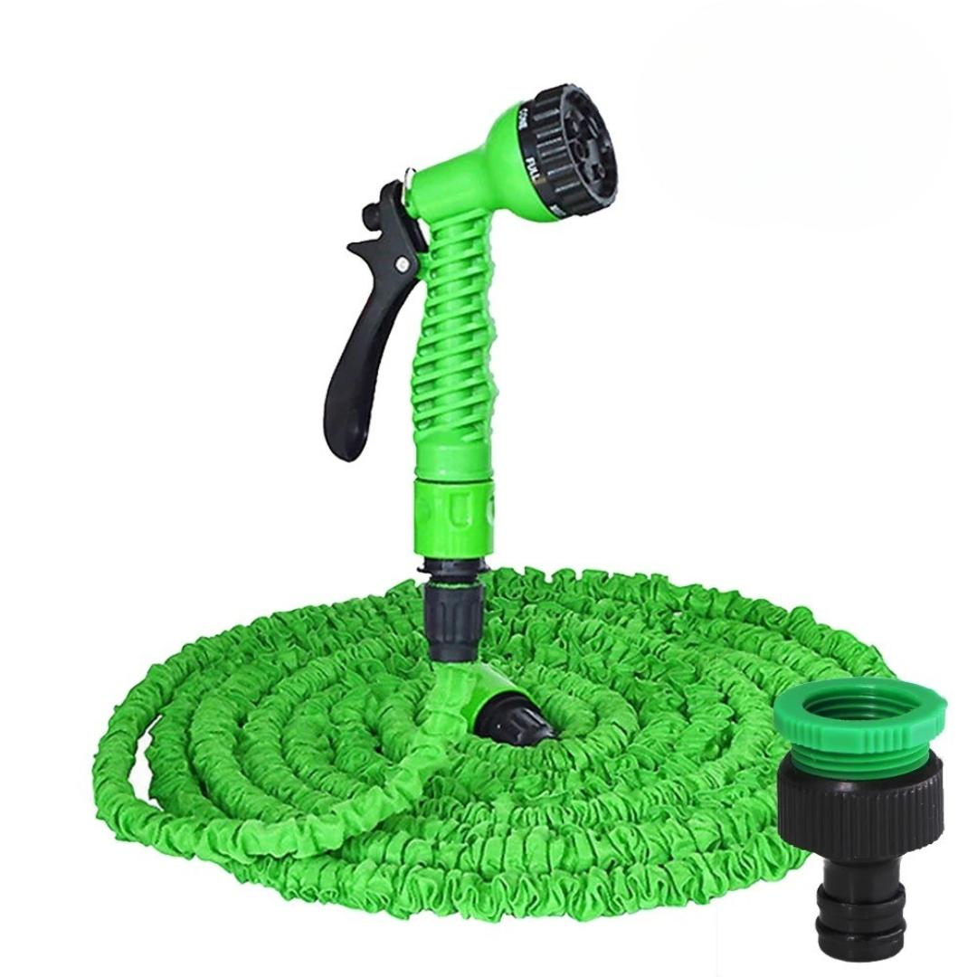
• Living Well with CAH: Challenges, Support, and Long-Term Outlook

    Living with Congenital Adrenal Hyperplasia presents unique challenges that require ongoing management, support, and a proactive approach to health. While CAH is a lifelong condition, with proper treatment and monitoring, individuals with CAH can lead healthy and fulfilling lives.
    https://www.marketresearchfuture.com/reports/congenital-adrenal-hyperplasia-market-4946

    One of the primary challenges for individuals with CAH and their families is the need for consistent hormone replacement therapy. Adherence to the prescribed medication regimen is crucial for maintaining hormonal balance and preventing potentially serious complications, such as adrenal crises. Education and understanding of the importance of medication, especially during times of stress or illness when cortisol needs may increase, are vital.

    Psychological and social support are also essential aspects of living with CAH. For individuals with ambiguous genitalia, the process of gender assignment and surgical correction can be emotionally challenging for both the child and the family. Open communication, counseling, and support groups can provide valuable resources and a sense of community. Individuals with NCAH may face challenges related to body image due to hirsutism and acne, as well as potential fertility issues, requiring sensitive and comprehensive care.

    Transitioning from pediatric to adult care is another critical period for individuals with CAH. Ensuring a smooth transfer of medical care to an adult endocrinologist who is knowledgeable about CAH is important for continued optimal management. Adult patients need ongoing education about their condition and the importance of lifelong adherence to treatment and monitoring.

    Potential long-term health considerations for individuals with CAH include the risk of adrenal crises if cortisol replacement is inadequate, potential metabolic issues related to glucocorticoid therapy (e.g., weight gain, bone density loss), and the long-term effects of androgen excess if not adequately suppressed. Regular monitoring and proactive management can help mitigate these risks.

    Support groups and patient advocacy organizations play a significant role in providing information, resources, and a sense of community for individuals with CAH and their families. Connecting with others who have similar experiences can be incredibly valuable for sharing coping strategies and navigating the challenges of living with a rare condition.

    The long-term outlook for individuals with CAH who receive appropriate and consistent medical care is generally good. With proper hormone replacement and regular monitoring, they can achieve normal growth and development, go through puberty, and lead active and healthy lives. Ongoing research continues to improve our understanding of CAH and develop new and more effective treatment strategies, offering hope for an even better future for those living with this condition.
    Living Well with CAH: Challenges, Support, and Long-Term Outlook Living with Congenital Adrenal Hyperplasia presents unique challenges that require ongoing management, support, and a proactive approach to health. While CAH is a lifelong condition, with proper treatment and monitoring, individuals with CAH can lead healthy and fulfilling lives. https://www.marketresearchfuture.com/reports/congenital-adrenal-hyperplasia-market-4946 One of the primary challenges for individuals with CAH and their families is the need for consistent hormone replacement therapy. Adherence to the prescribed medication regimen is crucial for maintaining hormonal balance and preventing potentially serious complications, such as adrenal crises. Education and understanding of the importance of medication, especially during times of stress or illness when cortisol needs may increase, are vital. Psychological and social support are also essential aspects of living with CAH. For individuals with ambiguous genitalia, the process of gender assignment and surgical correction can be emotionally challenging for both the child and the family. Open communication, counseling, and support groups can provide valuable resources and a sense of community. Individuals with NCAH may face challenges related to body image due to hirsutism and acne, as well as potential fertility issues, requiring sensitive and comprehensive care. Transitioning from pediatric to adult care is another critical period for individuals with CAH. Ensuring a smooth transfer of medical care to an adult endocrinologist who is knowledgeable about CAH is important for continued optimal management. Adult patients need ongoing education about their condition and the importance of lifelong adherence to treatment and monitoring. Potential long-term health considerations for individuals with CAH include the risk of adrenal crises if cortisol replacement is inadequate, potential metabolic issues related to glucocorticoid therapy (e.g., weight gain, bone density loss), and the long-term effects of androgen excess if not adequately suppressed. Regular monitoring and proactive management can help mitigate these risks. Support groups and patient advocacy organizations play a significant role in providing information, resources, and a sense of community for individuals with CAH and their families. Connecting with others who have similar experiences can be incredibly valuable for sharing coping strategies and navigating the challenges of living with a rare condition. The long-term outlook for individuals with CAH who receive appropriate and consistent medical care is generally good. With proper hormone replacement and regular monitoring, they can achieve normal growth and development, go through puberty, and lead active and healthy lives. Ongoing research continues to improve our understanding of CAH and develop new and more effective treatment strategies, offering hope for an even better future for those living with this condition.
    WWW.MARKETRESEARCHFUTURE.COM
    Congenital Adrenal Hyperplasia Market Size and Forecast to 2032
    Congenital Adrenal Hyperplasia Market is expected to grow at a CAGR over 6.79% By 2032. Congenital Adrenal Hyperplasia Market Analysis by Type, Treatment & Diagnosis, Treatment and End Users | Congenital Adrenal Hyperplasia Industry | MRFR
    0 Comments 0 Shares
  • The Dawn of a New Era: Understanding the Total Ankle Replacement Revolution

    For years, those suffering from debilitating ankle pain due to arthritis, injury, or other degenerative conditions often faced a limited set of solutions. Ankle fusion, while effective in eliminating pain, came at the cost of joint mobility, impacting gait and overall lifestyle. However, the landscape of ankle care is undergoing a significant transformation, a true revolution driven by advancements in total ankle replacement (TAR) technology and surgical techniques. This isn't just an incremental improvement; it's a paradigm shift offering the potential for pain relief and the preservation of natural ankle movement.

    https://www.marketresearchfuture.com/reports/total-ankle-replacement-market-42600

    The "revolution" in total ankle replacement stems from several key factors. Firstly, the design and materials used in ankle implants have evolved dramatically. Early generation implants faced challenges related to longevity and wear. Today, we see sophisticated designs incorporating durable materials like cobalt-chromium alloys and high-density polyethylene, engineered to withstand the complex biomechanical forces acting on the ankle joint. These advancements translate to improved implant lifespan and a reduced risk of revision surgery.

    Secondly, surgical techniques have become increasingly refined. Surgeons specializing in TAR are now utilizing minimally invasive approaches in some cases, leading to smaller incisions, reduced soft tissue damage, and potentially faster recovery times. Advanced imaging and computer-assisted navigation are also playing a growing role, enhancing surgical precision and implant alignment. This meticulous approach is crucial for optimizing implant function and minimizing the risk of complications.

    Furthermore, the understanding of ankle biomechanics has deepened considerably. Researchers and surgeons now have a more comprehensive grasp of the intricate movements and load distribution within the ankle joint. This knowledge informs the design of new implants that more closely mimic the natural kinematics of a healthy ankle, allowing for a smoother and more natural gait.

    The "revolution" also encompasses a growing awareness and acceptance of TAR as a viable alternative to fusion. As more patients experience successful outcomes and long-term relief, and as more data supports the efficacy of modern implants, both patients and referring physicians are increasingly considering TAR as a primary treatment option for end-stage ankle arthritis. This shift in perspective is crucial in empowering patients to explore a wider range of possibilities for regaining their mobility and quality of life.

    In essence, the total ankle replacement revolution is about offering patients a better future – one where chronic ankle pain doesn't dictate their limitations. It’s about combining cutting-edge technology, advanced surgical expertise, and a deeper understanding of the ankle to provide a solution that not only alleviates pain but also strives to restore natural function. As research continues and techniques evolve, this revolution promises to bring even greater hope and improved outcomes for individuals seeking to step into a pain-free tomorrow.
    The Dawn of a New Era: Understanding the Total Ankle Replacement Revolution For years, those suffering from debilitating ankle pain due to arthritis, injury, or other degenerative conditions often faced a limited set of solutions. Ankle fusion, while effective in eliminating pain, came at the cost of joint mobility, impacting gait and overall lifestyle. However, the landscape of ankle care is undergoing a significant transformation, a true revolution driven by advancements in total ankle replacement (TAR) technology and surgical techniques. This isn't just an incremental improvement; it's a paradigm shift offering the potential for pain relief and the preservation of natural ankle movement. https://www.marketresearchfuture.com/reports/total-ankle-replacement-market-42600 The "revolution" in total ankle replacement stems from several key factors. Firstly, the design and materials used in ankle implants have evolved dramatically. Early generation implants faced challenges related to longevity and wear. Today, we see sophisticated designs incorporating durable materials like cobalt-chromium alloys and high-density polyethylene, engineered to withstand the complex biomechanical forces acting on the ankle joint. These advancements translate to improved implant lifespan and a reduced risk of revision surgery. Secondly, surgical techniques have become increasingly refined. Surgeons specializing in TAR are now utilizing minimally invasive approaches in some cases, leading to smaller incisions, reduced soft tissue damage, and potentially faster recovery times. Advanced imaging and computer-assisted navigation are also playing a growing role, enhancing surgical precision and implant alignment. This meticulous approach is crucial for optimizing implant function and minimizing the risk of complications. Furthermore, the understanding of ankle biomechanics has deepened considerably. Researchers and surgeons now have a more comprehensive grasp of the intricate movements and load distribution within the ankle joint. This knowledge informs the design of new implants that more closely mimic the natural kinematics of a healthy ankle, allowing for a smoother and more natural gait. The "revolution" also encompasses a growing awareness and acceptance of TAR as a viable alternative to fusion. As more patients experience successful outcomes and long-term relief, and as more data supports the efficacy of modern implants, both patients and referring physicians are increasingly considering TAR as a primary treatment option for end-stage ankle arthritis. This shift in perspective is crucial in empowering patients to explore a wider range of possibilities for regaining their mobility and quality of life. In essence, the total ankle replacement revolution is about offering patients a better future – one where chronic ankle pain doesn't dictate their limitations. It’s about combining cutting-edge technology, advanced surgical expertise, and a deeper understanding of the ankle to provide a solution that not only alleviates pain but also strives to restore natural function. As research continues and techniques evolve, this revolution promises to bring even greater hope and improved outcomes for individuals seeking to step into a pain-free tomorrow.
    WWW.MARKETRESEARCHFUTURE.COM
    Total Ankle Replacement Market Size, Share, Trends 2035 | MRFR
    Total Ankle Replacement Market 2025, Industry to reach USD 2.15 Billion, at a 6.46% CAGR by growing size, share, top company analysis, segments research, trends and forecast to 2035
    0 Comments 0 Shares
  • Considering a Partial Knee Replacement? What You Need to Know About Unicompartmental Knee Prosthesis

    If you're experiencing persistent knee pain, particularly localized to one side of your knee, and traditional conservative treatments like physical therapy, medication, and injections are no longer providing adequate relief, your orthopedic surgeon may have discussed the possibility of a partial knee replacement, also known as a unicompartmental knee prosthesis. This surgical option offers a less invasive alternative to a total knee replacement for individuals with arthritis that primarily affects only one compartment of the knee. Understanding the nuances of a unicompartmental knee prosthesis – what it entails, its benefits, the eligibility criteria, and what to expect during recovery – is crucial for making an informed decision about your treatment journey and taking the first steps towards regaining pain-free mobility.

    https://www.marketresearchfuture.com/reports/unicompartmental-knee-prosthese-market-40253

    The knee joint is comprised of three main compartments: the medial (inner) compartment, the lateral (outer) compartment, and the patellofemoral compartment (behind the kneecap). Osteoarthritis, the most common reason for knee replacement surgery, often affects only one or two of these compartments. In contrast to a total knee replacement, where all three compartments are resurfaced and replaced with prosthetic components, a unicompartmental knee replacement focuses solely on the damaged compartment. This less extensive approach offers several potential advantages, which we will explore further.

    During a unicompartmental knee replacement procedure, the damaged cartilage and bone in the affected compartment are removed and replaced with a smooth metal and plastic prosthesis. The healthy cartilage and bone in the remaining compartments are left intact. This targeted intervention preserves more of the knee's natural anatomy, which can contribute to a more natural feeling knee after surgery and potentially a greater range of motion compared to a total knee replacement in some individuals.

    The prosthetic components used in a unicompartmental knee replacement typically consist of a metal femoral component that resurfaces the end of the thigh bone (femur) in the affected compartment and a tibial component, usually a metal tray with a smooth plastic bearing surface, that replaces the top of the shin bone (tibia) in the same compartment. These components are carefully sized and positioned to restore the smooth gliding motion of the knee joint and alleviate pain caused by bone-on-bone friction in the damaged area.

    The surgical procedure for a unicompartmental knee replacement is generally less complex and invasive than a total knee replacement. This often translates to a smaller incision, less blood loss, and a shorter hospital stay. The reduced surgical trauma can also contribute to a faster and less painful recovery process for many patients.

    However, it's important to understand that a unicompartmental knee replacement is not suitable for everyone with knee arthritis. Specific criteria must be met to be considered a good candidate for this procedure. These criteria typically include significant pain primarily localized to one compartment of the knee, relatively good range of motion before surgery, stable ligaments, and minimal deformity of the knee. If arthritis affects multiple compartments or if there is significant ligament instability or knee deformity, a total knee replacement may be a more appropriate option.

    If you are considering a partial knee replacement, a thorough evaluation by an experienced orthopedic surgeon is essential. This evaluation will involve a detailed medical history, a physical examination of your knee, and imaging studies such as X-rays and potentially MRI scans to assess the extent and location of the arthritis. Based on this comprehensive assessment, your surgeon will determine if a unicompartmental knee prosthesis is the right treatment option to address your specific needs and help you return to a more active and pain-free lifestyle. Open communication with your surgeon, asking questions, and understanding the potential benefits and risks are key steps in making an informed decision about your knee care.
    Considering a Partial Knee Replacement? What You Need to Know About Unicompartmental Knee Prosthesis If you're experiencing persistent knee pain, particularly localized to one side of your knee, and traditional conservative treatments like physical therapy, medication, and injections are no longer providing adequate relief, your orthopedic surgeon may have discussed the possibility of a partial knee replacement, also known as a unicompartmental knee prosthesis. This surgical option offers a less invasive alternative to a total knee replacement for individuals with arthritis that primarily affects only one compartment of the knee. Understanding the nuances of a unicompartmental knee prosthesis – what it entails, its benefits, the eligibility criteria, and what to expect during recovery – is crucial for making an informed decision about your treatment journey and taking the first steps towards regaining pain-free mobility. https://www.marketresearchfuture.com/reports/unicompartmental-knee-prosthese-market-40253 The knee joint is comprised of three main compartments: the medial (inner) compartment, the lateral (outer) compartment, and the patellofemoral compartment (behind the kneecap). Osteoarthritis, the most common reason for knee replacement surgery, often affects only one or two of these compartments. In contrast to a total knee replacement, where all three compartments are resurfaced and replaced with prosthetic components, a unicompartmental knee replacement focuses solely on the damaged compartment. This less extensive approach offers several potential advantages, which we will explore further. During a unicompartmental knee replacement procedure, the damaged cartilage and bone in the affected compartment are removed and replaced with a smooth metal and plastic prosthesis. The healthy cartilage and bone in the remaining compartments are left intact. This targeted intervention preserves more of the knee's natural anatomy, which can contribute to a more natural feeling knee after surgery and potentially a greater range of motion compared to a total knee replacement in some individuals. The prosthetic components used in a unicompartmental knee replacement typically consist of a metal femoral component that resurfaces the end of the thigh bone (femur) in the affected compartment and a tibial component, usually a metal tray with a smooth plastic bearing surface, that replaces the top of the shin bone (tibia) in the same compartment. These components are carefully sized and positioned to restore the smooth gliding motion of the knee joint and alleviate pain caused by bone-on-bone friction in the damaged area. The surgical procedure for a unicompartmental knee replacement is generally less complex and invasive than a total knee replacement. This often translates to a smaller incision, less blood loss, and a shorter hospital stay. The reduced surgical trauma can also contribute to a faster and less painful recovery process for many patients. However, it's important to understand that a unicompartmental knee replacement is not suitable for everyone with knee arthritis. Specific criteria must be met to be considered a good candidate for this procedure. These criteria typically include significant pain primarily localized to one compartment of the knee, relatively good range of motion before surgery, stable ligaments, and minimal deformity of the knee. If arthritis affects multiple compartments or if there is significant ligament instability or knee deformity, a total knee replacement may be a more appropriate option. If you are considering a partial knee replacement, a thorough evaluation by an experienced orthopedic surgeon is essential. This evaluation will involve a detailed medical history, a physical examination of your knee, and imaging studies such as X-rays and potentially MRI scans to assess the extent and location of the arthritis. Based on this comprehensive assessment, your surgeon will determine if a unicompartmental knee prosthesis is the right treatment option to address your specific needs and help you return to a more active and pain-free lifestyle. Open communication with your surgeon, asking questions, and understanding the potential benefits and risks are key steps in making an informed decision about your knee care.
    WWW.MARKETRESEARCHFUTURE.COM
    Unicompartmental Knee Prosthese Market Size, Share, Growth Report 2034
    Unicompartmental Knee Prosthese Market growth is projected to reach USD 5.32 Billion, at a 5.66% CAGR by driving industry size, share, top company analysis, segments research, trends and forecast report 2025 to 2034
    0 Comments 0 Shares
  • The World of Americas Acrylic Teeth: A Foundation for Countless Smiles

    In the realm of dental prosthetics across the Americas, acrylic teeth stand as a cornerstone, providing an affordable and aesthetically pleasing solution for replacing missing teeth. From full dentures to partial replacements, these artificial teeth, primarily made from polymethyl methacrylate (PMMA), play a vital role in restoring smiles, function, and confidence for millions.

    Acrylic teeth have gained immense popularity due to several key advantages:

    Cost-Effectiveness: Compared to porcelain or other materials, acrylic teeth offer a more budget-friendly option for patients seeking tooth replacement. This affordability makes them accessible to a wider population.

    Aesthetics: Modern acrylic resins can be expertly crafted to mimic the natural shade, shape, and translucency of real teeth and gums. Skilled technicians can achieve highly realistic results, ensuring a natural-looking smile.

    Comfort: Being lighter than porcelain, acrylic dentures tend to be more comfortable for many wearers, especially during the initial adjustment period.

    Ease of Adjustment: Dentists and lab technicians can easily adjust and modify acrylic dentures to ensure a proper and comfortable fit.

    Bonding Ability: Acrylic teeth chemically bond well with the acrylic denture base, creating a strong and integrated prosthesis.

    Reduced Wear on Natural Teeth: In partial dentures, acrylic teeth exert less abrasive force on the remaining natural teeth compared to some other materials.

    Versatility: Acrylic teeth are suitable for both complete and partial dentures, as well as overdentures.

    The market for acrylic teeth in the Americas is significant and continues to grow, driven by factors such as an aging population, increasing awareness of oral health, and advancements in dental materials and manufacturing technologies like 3D printing. This makes the "Americas Acrylic Teeth" market a dynamic and important sector within the broader dental industry.
    The World of Americas Acrylic Teeth: A Foundation for Countless Smiles In the realm of dental prosthetics across the Americas, acrylic teeth stand as a cornerstone, providing an affordable and aesthetically pleasing solution for replacing missing teeth. From full dentures to partial replacements, these artificial teeth, primarily made from polymethyl methacrylate (PMMA), play a vital role in restoring smiles, function, and confidence for millions. Acrylic teeth have gained immense popularity due to several key advantages: Cost-Effectiveness: Compared to porcelain or other materials, acrylic teeth offer a more budget-friendly option for patients seeking tooth replacement. This affordability makes them accessible to a wider population. Aesthetics: Modern acrylic resins can be expertly crafted to mimic the natural shade, shape, and translucency of real teeth and gums. Skilled technicians can achieve highly realistic results, ensuring a natural-looking smile. Comfort: Being lighter than porcelain, acrylic dentures tend to be more comfortable for many wearers, especially during the initial adjustment period. Ease of Adjustment: Dentists and lab technicians can easily adjust and modify acrylic dentures to ensure a proper and comfortable fit. Bonding Ability: Acrylic teeth chemically bond well with the acrylic denture base, creating a strong and integrated prosthesis. Reduced Wear on Natural Teeth: In partial dentures, acrylic teeth exert less abrasive force on the remaining natural teeth compared to some other materials. Versatility: Acrylic teeth are suitable for both complete and partial dentures, as well as overdentures. The market for acrylic teeth in the Americas is significant and continues to grow, driven by factors such as an aging population, increasing awareness of oral health, and advancements in dental materials and manufacturing technologies like 3D printing. This makes the "Americas Acrylic Teeth" market a dynamic and important sector within the broader dental industry.
    0 Comments 0 Shares
  • The Crucial Treatment: Relieving the Pressure in Cardiac Tamponade

    Cardiac tamponade is a medical emergency that requires immediate intervention to relieve the pressure on the heart and restore 1 its ability to pump blood effectively. The primary treatment strategy involves removing the excess fluid from the pericardial sac.  
    https://www.marketresearchfuture.com/reports/cardiac-tamponade-market-4341

    Emergency Measures:

    In the initial management of a patient with suspected cardiac tamponade, supportive measures are crucial:

    Oxygen Administration: To help improve oxygenation to the tissues.
    Intravenous (IV) Fluids: To increase preload (the amount of blood filling the heart) and help maintain blood pressure until the fluid can be removed.

    Vasopressors: Medications to help increase blood pressure may be used temporarily.

    Close Monitoring: Continuous monitoring of vital signs (heart rate, blood pressure, oxygen saturation) is essential.

    Definitive Treatment: Pericardiocentesis:The most common and often the first-line definitive treatment for cardiac tamponade is pericardiocentesis. This is a minimally invasive procedure performed by inserting a needle into the chest wall and guiding it (often with the aid of echocardiography) into the pericardial sac to drain the accumulated fluid.

    The steps involved in pericardiocentesis typically include:

    Preparation: The patient's skin is cleaned and numbed with local anesthesia.

    Needle Insertion: A long needle is carefully inserted into the pericardial space, usually just below the rib cage on the left side or through another designated entry point guided by imaging.

    Fluid Drainage: Once the needle is in the pericardial sac, fluid is withdrawn using a syringe.

    Catheter Placement (Optional): In some cases, a thin plastic tube (catheter) may be left in place for continuous drainage of fluid over several hours or days, especially if the effusion is large or likely to reaccumulate.

    Pericardiocentesis can provide rapid relief of the pressure on the heart, leading to immediate improvement in blood pressure and cardiac output. The drained fluid is often sent to the laboratory for analysis to help determine the underlying cause of the tamponade.

    Surgical Intervention:
    In certain situations, surgical intervention may be necessary to treat cardiac tamponade:
    Thoracotomy or Pericardial Window: If pericardiocentesis is unsuccessful, if there is recurrent tamponade, or if the cause is related to trauma (e.g., bleeding from a heart laceration) or certain complications of heart surgery, a surgical approach may be required. This involves opening the chest (thoracotomy) or creating a "window" in the pericardium (pericardial window) to allow the fluid to drain into the pleural space or abdomen, where it can be absorbed.
    Repair of Underlying Cause: Surgery may also be needed to address the underlying cause of the tamponade, such as repairing a heart rupture after a myocardial infarction or addressing an aortic dissection.

    Pericardiectomy: In cases of chronic recurrent pericardial effusion leading to tamponade, surgical removal of part or all of the pericardium (pericardiectomy) may be considered to prevent further fluid accumulation and constriction of the heart.

    Treating the Underlying Cause:
    It's crucial to remember that while relieving the immediate pressure on the heart is the priority, identifying and treating the underlying cause of the cardiac tamponade is equally important for long-term management and preventing recurrence. For example, if the tamponade is due to an infection, antibiotics will be necessary. If it's due to cancer, treatment for the malignancy will be required.

    The rapid recognition and appropriate treatment of cardiac tamponade are critical for improving patient outcomes and saving lives.
    The Crucial Treatment: Relieving the Pressure in Cardiac Tamponade Cardiac tamponade is a medical emergency that requires immediate intervention to relieve the pressure on the heart and restore 1 its ability to pump blood effectively. The primary treatment strategy involves removing the excess fluid from the pericardial sac.   https://www.marketresearchfuture.com/reports/cardiac-tamponade-market-4341 Emergency Measures: In the initial management of a patient with suspected cardiac tamponade, supportive measures are crucial: Oxygen Administration: To help improve oxygenation to the tissues. Intravenous (IV) Fluids: To increase preload (the amount of blood filling the heart) and help maintain blood pressure until the fluid can be removed. Vasopressors: Medications to help increase blood pressure may be used temporarily. Close Monitoring: Continuous monitoring of vital signs (heart rate, blood pressure, oxygen saturation) is essential. Definitive Treatment: Pericardiocentesis:The most common and often the first-line definitive treatment for cardiac tamponade is pericardiocentesis. This is a minimally invasive procedure performed by inserting a needle into the chest wall and guiding it (often with the aid of echocardiography) into the pericardial sac to drain the accumulated fluid. The steps involved in pericardiocentesis typically include: Preparation: The patient's skin is cleaned and numbed with local anesthesia. Needle Insertion: A long needle is carefully inserted into the pericardial space, usually just below the rib cage on the left side or through another designated entry point guided by imaging. Fluid Drainage: Once the needle is in the pericardial sac, fluid is withdrawn using a syringe. Catheter Placement (Optional): In some cases, a thin plastic tube (catheter) may be left in place for continuous drainage of fluid over several hours or days, especially if the effusion is large or likely to reaccumulate. Pericardiocentesis can provide rapid relief of the pressure on the heart, leading to immediate improvement in blood pressure and cardiac output. The drained fluid is often sent to the laboratory for analysis to help determine the underlying cause of the tamponade. Surgical Intervention: In certain situations, surgical intervention may be necessary to treat cardiac tamponade: Thoracotomy or Pericardial Window: If pericardiocentesis is unsuccessful, if there is recurrent tamponade, or if the cause is related to trauma (e.g., bleeding from a heart laceration) or certain complications of heart surgery, a surgical approach may be required. This involves opening the chest (thoracotomy) or creating a "window" in the pericardium (pericardial window) to allow the fluid to drain into the pleural space or abdomen, where it can be absorbed. Repair of Underlying Cause: Surgery may also be needed to address the underlying cause of the tamponade, such as repairing a heart rupture after a myocardial infarction or addressing an aortic dissection. Pericardiectomy: In cases of chronic recurrent pericardial effusion leading to tamponade, surgical removal of part or all of the pericardium (pericardiectomy) may be considered to prevent further fluid accumulation and constriction of the heart. Treating the Underlying Cause: It's crucial to remember that while relieving the immediate pressure on the heart is the priority, identifying and treating the underlying cause of the cardiac tamponade is equally important for long-term management and preventing recurrence. For example, if the tamponade is due to an infection, antibiotics will be necessary. If it's due to cancer, treatment for the malignancy will be required. The rapid recognition and appropriate treatment of cardiac tamponade are critical for improving patient outcomes and saving lives.
    DoveMed | Trusted health information for a better life
    Trusted health information for a better life on health, wellness, diseases, surgical and radiology procedures, lab tests, first aid, nutrition and green living.
    0 Comments 0 Shares
  • Understanding Hypoprothrombinemia: Causes, Symptoms, and Effective Treatments

    Hypoprothrombinemia is a rare but serious bleeding disorder characterized by abnormally low levels of prothrombin, a vital protein involved in blood clotting. Without sufficient prothrombin, the blood cannot clot properly, which may lead to spontaneous bleeding or difficulty stopping bleeding after injury. This condition can arise due to genetic factors or as an acquired disorder and demands timely diagnosis and treatment.

    The most common cause of hypoprothrombinemia is a deficiency in vitamin K. Vitamin K is crucial for synthesizing prothrombin in the liver. When the body doesn’t get enough vitamin K—either through poor diet, malabsorption syndromes (like celiac or Crohn’s disease), or prolonged antibiotic use—prothrombin levels can drop. Another cause can be liver disease, since the liver is responsible for producing clotting factors.

    https://www.marketresearchfuture.com/reports/hypoprothrombinemia-treatment-market-37949

    There’s also a rarer, inherited form of hypoprothrombinemia known as congenital hypoprothrombinemia, which occurs due to mutations in the F2 gene. This genetic variation affects the body's ability to produce normal levels of prothrombin. Symptoms typically appear in childhood and include frequent nosebleeds, easy bruising, and prolonged bleeding from cuts or surgical procedures.

    Symptoms of hypoprothrombinemia vary depending on severity. Mild cases may go unnoticed until a bleeding event occurs. More serious cases involve spontaneous internal bleeding, joint swelling due to bleeding into joints (hemarthrosis), or gastrointestinal bleeding. In newborns, the condition may present as bleeding from the umbilical stump, gastrointestinal tract, or even the brain if left untreated.

    Diagnosis usually involves blood tests such as prothrombin time (PT), which measures how long it takes blood to clot. A prolonged PT may indicate low prothrombin levels. Additional tests, including vitamin K levels, liver function tests, and genetic studies, help pinpoint the underlying cause.

    Treatment of hypoprothrombinemia largely depends on the cause. In cases related to vitamin K deficiency, the solution is relatively straightforward: vitamin K supplementation. This can be given orally or via injection, especially in emergencies where rapid correction is needed. Most patients respond quickly to vitamin K therapy with normalization of clotting times.

    If the condition is caused by liver disease, treatment focuses on managing the liver issue alongside administering vitamin K. In more severe cases, plasma transfusions or prothrombin complex concentrates (PCCs) may be necessary to restore clotting function quickly. These are especially useful during surgeries or active bleeding episodes.

    For congenital hypoprothrombinemia, regular monitoring and prophylactic treatment may be required. In such cases, replacement therapy with fresh frozen plasma or factor concentrates is typically used before surgical or dental procedures to prevent bleeding complications.

    Early detection is crucial to prevent severe complications. Regular monitoring of clotting function and liver health is important, especially in high-risk individuals. Infants are commonly given a vitamin K injection at birth to prevent bleeding disorders, including hypoprothrombinemia.

    In summary, hypoprothrombinemia is a potentially serious bleeding disorder that requires accurate diagnosis and appropriate treatment. Whether caused by vitamin K deficiency, liver dysfunction, or a genetic mutation, effective therapies are available to restore prothrombin levels and prevent bleeding. With proper medical care and ongoing monitoring, most individuals with this condition can lead normal, healthy lives.
    Understanding Hypoprothrombinemia: Causes, Symptoms, and Effective Treatments Hypoprothrombinemia is a rare but serious bleeding disorder characterized by abnormally low levels of prothrombin, a vital protein involved in blood clotting. Without sufficient prothrombin, the blood cannot clot properly, which may lead to spontaneous bleeding or difficulty stopping bleeding after injury. This condition can arise due to genetic factors or as an acquired disorder and demands timely diagnosis and treatment. The most common cause of hypoprothrombinemia is a deficiency in vitamin K. Vitamin K is crucial for synthesizing prothrombin in the liver. When the body doesn’t get enough vitamin K—either through poor diet, malabsorption syndromes (like celiac or Crohn’s disease), or prolonged antibiotic use—prothrombin levels can drop. Another cause can be liver disease, since the liver is responsible for producing clotting factors. https://www.marketresearchfuture.com/reports/hypoprothrombinemia-treatment-market-37949 There’s also a rarer, inherited form of hypoprothrombinemia known as congenital hypoprothrombinemia, which occurs due to mutations in the F2 gene. This genetic variation affects the body's ability to produce normal levels of prothrombin. Symptoms typically appear in childhood and include frequent nosebleeds, easy bruising, and prolonged bleeding from cuts or surgical procedures. Symptoms of hypoprothrombinemia vary depending on severity. Mild cases may go unnoticed until a bleeding event occurs. More serious cases involve spontaneous internal bleeding, joint swelling due to bleeding into joints (hemarthrosis), or gastrointestinal bleeding. In newborns, the condition may present as bleeding from the umbilical stump, gastrointestinal tract, or even the brain if left untreated. Diagnosis usually involves blood tests such as prothrombin time (PT), which measures how long it takes blood to clot. A prolonged PT may indicate low prothrombin levels. Additional tests, including vitamin K levels, liver function tests, and genetic studies, help pinpoint the underlying cause. Treatment of hypoprothrombinemia largely depends on the cause. In cases related to vitamin K deficiency, the solution is relatively straightforward: vitamin K supplementation. This can be given orally or via injection, especially in emergencies where rapid correction is needed. Most patients respond quickly to vitamin K therapy with normalization of clotting times. If the condition is caused by liver disease, treatment focuses on managing the liver issue alongside administering vitamin K. In more severe cases, plasma transfusions or prothrombin complex concentrates (PCCs) may be necessary to restore clotting function quickly. These are especially useful during surgeries or active bleeding episodes. For congenital hypoprothrombinemia, regular monitoring and prophylactic treatment may be required. In such cases, replacement therapy with fresh frozen plasma or factor concentrates is typically used before surgical or dental procedures to prevent bleeding complications. Early detection is crucial to prevent severe complications. Regular monitoring of clotting function and liver health is important, especially in high-risk individuals. Infants are commonly given a vitamin K injection at birth to prevent bleeding disorders, including hypoprothrombinemia. In summary, hypoprothrombinemia is a potentially serious bleeding disorder that requires accurate diagnosis and appropriate treatment. Whether caused by vitamin K deficiency, liver dysfunction, or a genetic mutation, effective therapies are available to restore prothrombin levels and prevent bleeding. With proper medical care and ongoing monitoring, most individuals with this condition can lead normal, healthy lives.
    WWW.MARKETRESEARCHFUTURE.COM
    Hypoprothrombinemia Treatment Market Size, Share, Growth Report 2034
    Hypoprothrombinemia Treatment Market growth is projected to reach USD 1.78 Billion, at a 6.04% CAGR by driving industry size, share, top company analysis, segments research, trends and forecast report 2025 to 2034.
    0 Comments 0 Shares
  • Innovative Hybrid Solutions Combine C-Arm and Portable X-ray Functionality: New Devices are Merging the Benefits of Both C-arms and Portable X-ray Systems.

    Recognizing the complementary strengths of C-arm and portable X-ray systems, innovative manufacturers are developing hybrid solutions that seamlessly integrate the functionalities of both technologies into a single, versatile device. These new hybrid systems aim to provide healthcare facilities with a cost-effective and space-saving solution that can address a wider range of imaging needs, merging the real-time fluoroscopic capabilities of C-arms with the convenience and portability of mobile X-ray units.

    https://www.marketresearchfuture.com/reports/c-arms-devices-market-17178

    Traditional C-arms excel at providing continuous, real-time fluoroscopic imaging, which is essential for guiding interventional procedures and visualizing dynamic processes. Portable X-ray systems, on the other hand, offer the advantage of bringing static radiographic imaging directly to the patient's bedside, particularly for patients who are too ill or immobile to be transported to a radiology department. Hybrid solutions seek to combine these distinct benefits into a single platform.

    One of the key features of these innovative hybrid solutions is their ability to switch between fluoroscopic and radiographic imaging modes. This allows clinicians to perform both dynamic and static imaging studies with the same device, eliminating the need to move patients between different imaging systems or bring in multiple pieces of equipment. For instance, a hybrid system could be used for real-time guidance during the placement of a central line in the intensive care unit and then immediately utilized to obtain a chest X-ray to confirm placement, all at the patient's bedside.

    The design of these hybrid devices often incorporates features that facilitate both types of imaging. They typically include a C-arm gantry for maneuverability and positioning during fluoroscopy, along with a powerful X-ray generator and detector capable of producing high-quality radiographic images. Some hybrid systems may also feature specialized software and workflows optimized for both fluoroscopic and radiographic applications.

    The potential benefits of these hybrid solutions are numerous. They can lead to cost savings by reducing the need to purchase and maintain separate C-arm and portable X-ray units. They can also save space in crowded healthcare environments by consolidating two imaging functionalities into a single device. Furthermore, they can improve workflow efficiency by allowing clinicians to perform a wider range of imaging studies with a single, readily available system, minimizing patient transport and delays.

    Enhanced patient convenience and safety are also potential advantages of hybrid solutions. By bringing the imaging directly to the patient, especially for bedside radiography, the risks associated with patient transport are reduced. The ability to perform both fluoroscopy and radiography without moving the patient can also improve comfort and streamline the diagnostic and interventional process.

    The expanding applications of these hybrid devices are significant. They can be utilized in emergency departments for rapid assessment of trauma patients, in intensive care units for bedside imaging and procedural guidance, in operating rooms for intraoperative fluoroscopy and post-operative radiographs, and in various other clinical settings where both dynamic and static imaging are frequently required.

    While still a relatively emerging technology, innovative hybrid solutions combining C-arm and portable X-ray functionality represent a promising direction in medical imaging. By merging the strengths of these two essential modalities, these devices offer increased versatility, efficiency, and potential cost savings for healthcare facilities, ultimately contributing to improved patient care. As technology continues to advance, we can expect to see further development and adoption of these integrated imaging platforms.
    Innovative Hybrid Solutions Combine C-Arm and Portable X-ray Functionality: New Devices are Merging the Benefits of Both C-arms and Portable X-ray Systems. Recognizing the complementary strengths of C-arm and portable X-ray systems, innovative manufacturers are developing hybrid solutions that seamlessly integrate the functionalities of both technologies into a single, versatile device. These new hybrid systems aim to provide healthcare facilities with a cost-effective and space-saving solution that can address a wider range of imaging needs, merging the real-time fluoroscopic capabilities of C-arms with the convenience and portability of mobile X-ray units. https://www.marketresearchfuture.com/reports/c-arms-devices-market-17178 Traditional C-arms excel at providing continuous, real-time fluoroscopic imaging, which is essential for guiding interventional procedures and visualizing dynamic processes. Portable X-ray systems, on the other hand, offer the advantage of bringing static radiographic imaging directly to the patient's bedside, particularly for patients who are too ill or immobile to be transported to a radiology department. Hybrid solutions seek to combine these distinct benefits into a single platform. One of the key features of these innovative hybrid solutions is their ability to switch between fluoroscopic and radiographic imaging modes. This allows clinicians to perform both dynamic and static imaging studies with the same device, eliminating the need to move patients between different imaging systems or bring in multiple pieces of equipment. For instance, a hybrid system could be used for real-time guidance during the placement of a central line in the intensive care unit and then immediately utilized to obtain a chest X-ray to confirm placement, all at the patient's bedside. The design of these hybrid devices often incorporates features that facilitate both types of imaging. They typically include a C-arm gantry for maneuverability and positioning during fluoroscopy, along with a powerful X-ray generator and detector capable of producing high-quality radiographic images. Some hybrid systems may also feature specialized software and workflows optimized for both fluoroscopic and radiographic applications. The potential benefits of these hybrid solutions are numerous. They can lead to cost savings by reducing the need to purchase and maintain separate C-arm and portable X-ray units. They can also save space in crowded healthcare environments by consolidating two imaging functionalities into a single device. Furthermore, they can improve workflow efficiency by allowing clinicians to perform a wider range of imaging studies with a single, readily available system, minimizing patient transport and delays. Enhanced patient convenience and safety are also potential advantages of hybrid solutions. By bringing the imaging directly to the patient, especially for bedside radiography, the risks associated with patient transport are reduced. The ability to perform both fluoroscopy and radiography without moving the patient can also improve comfort and streamline the diagnostic and interventional process. The expanding applications of these hybrid devices are significant. They can be utilized in emergency departments for rapid assessment of trauma patients, in intensive care units for bedside imaging and procedural guidance, in operating rooms for intraoperative fluoroscopy and post-operative radiographs, and in various other clinical settings where both dynamic and static imaging are frequently required. While still a relatively emerging technology, innovative hybrid solutions combining C-arm and portable X-ray functionality represent a promising direction in medical imaging. By merging the strengths of these two essential modalities, these devices offer increased versatility, efficiency, and potential cost savings for healthcare facilities, ultimately contributing to improved patient care. As technology continues to advance, we can expect to see further development and adoption of these integrated imaging platforms.
    WWW.MARKETRESEARCHFUTURE.COM
    C Arms Devices Market Size, Growth, Trends, Report 2035
    C Arms Devices Market projected to grow at 5.3% CAGR, reaching USD 4 Billion by 2035, driving growth global trends, competitive industry analysis and outlook 2025-2035
    0 Comments 0 Shares
  • Pediatric Spinal Needles: Design Considerations and Techniques for Safe and Effective Use in Children

    Performing neuraxial procedures in children presents unique challenges due to their smaller size, differing anatomy, and increased vulnerability. The design of pediatric spinal needles and the techniques employed for their use must be carefully tailored to ensure safety, minimize trauma, and achieve effective anesthesia or diagnostic sampling in this sensitive population.
    https://www.marketresearchfuture.com/reports/spinal-needles-market-1216
    Needle size (gauge) and length are critical design considerations for pediatric spinal needles. Compared to adult needles, pediatric needles are typically smaller in diameter to minimize the risk of dural puncture and associated complications in smaller spinal canals. The length of the needle must also be appropriate for the child's age and size to ensure adequate penetration without excessive depth. A range of pediatric needle sizes and lengths are available to accommodate the diverse needs of this patient population, from neonates to adolescents.

    Needle tip design is also important. While atraumatic pencil-point needles have shown benefits in reducing PDPH in adults, their use in pediatrics is still evolving. The smaller size and potentially different dural characteristics in children require careful consideration of the optimal needle tip geometry to balance ease of insertion with minimal dural trauma. Some pediatric-specific needles incorporate modified bevel designs aimed at atraumatic puncture in this age group.

    Safety features are paramount in pediatric spinal needles. Features such as depth markings on the needle shaft can help guide insertion and prevent over-insertion. Some needles also incorporate mechanisms to provide tactile or visual confirmation of successful subarachnoid space entry, minimizing the risk of traumatic taps.  

    Techniques for spinal needle insertion in children often differ from those used in adults. Positioning is crucial and may require specialized aids to ensure the child is comfortable and still. Gentle and slow needle advancement is essential to minimize trauma. The "loss of resistance" technique, commonly used in adults for epidural placement, can be challenging to interpret in small children due to the shorter distances and less distinct tissue layers.  

    Ultrasound guidance plays an increasingly important role in pediatric spinal needle placement. The ability to visualize the spinal anatomy in real-time can be particularly beneficial in infants and young children where bony landmarks may be less easily palpable. Ultrasound can help identify the optimal insertion point, estimate the depth to the subarachnoid space, and guide needle advancement, improving first-pass success and reducing the risk of complications.

    Aseptic technique is even more critical in children due to their developing immune systems. Strict adherence to sterile protocols during preparation and the procedure is essential to minimize the risk of infection.  

    The choice of needle size, design, and insertion technique in pediatric spinal anesthesia and lumbar puncture requires careful consideration of the child's age, size, clinical condition, and the practitioner's experience. Ongoing research and the development of pediatric-specific spinal needles and techniques are crucial for ensuring the safe and effective use of neuraxial procedures in this vulnerable population. A focus on minimizing trauma, ensuring accurate placement, and employing strategies like ultrasound guidance are essential for optimizing outcomes and providing the best possible care for children undergoing these procedures.
    Pediatric Spinal Needles: Design Considerations and Techniques for Safe and Effective Use in Children Performing neuraxial procedures in children presents unique challenges due to their smaller size, differing anatomy, and increased vulnerability. The design of pediatric spinal needles and the techniques employed for their use must be carefully tailored to ensure safety, minimize trauma, and achieve effective anesthesia or diagnostic sampling in this sensitive population. https://www.marketresearchfuture.com/reports/spinal-needles-market-1216 Needle size (gauge) and length are critical design considerations for pediatric spinal needles. Compared to adult needles, pediatric needles are typically smaller in diameter to minimize the risk of dural puncture and associated complications in smaller spinal canals. The length of the needle must also be appropriate for the child's age and size to ensure adequate penetration without excessive depth. A range of pediatric needle sizes and lengths are available to accommodate the diverse needs of this patient population, from neonates to adolescents. Needle tip design is also important. While atraumatic pencil-point needles have shown benefits in reducing PDPH in adults, their use in pediatrics is still evolving. The smaller size and potentially different dural characteristics in children require careful consideration of the optimal needle tip geometry to balance ease of insertion with minimal dural trauma. Some pediatric-specific needles incorporate modified bevel designs aimed at atraumatic puncture in this age group. Safety features are paramount in pediatric spinal needles. Features such as depth markings on the needle shaft can help guide insertion and prevent over-insertion. Some needles also incorporate mechanisms to provide tactile or visual confirmation of successful subarachnoid space entry, minimizing the risk of traumatic taps.   Techniques for spinal needle insertion in children often differ from those used in adults. Positioning is crucial and may require specialized aids to ensure the child is comfortable and still. Gentle and slow needle advancement is essential to minimize trauma. The "loss of resistance" technique, commonly used in adults for epidural placement, can be challenging to interpret in small children due to the shorter distances and less distinct tissue layers.   Ultrasound guidance plays an increasingly important role in pediatric spinal needle placement. The ability to visualize the spinal anatomy in real-time can be particularly beneficial in infants and young children where bony landmarks may be less easily palpable. Ultrasound can help identify the optimal insertion point, estimate the depth to the subarachnoid space, and guide needle advancement, improving first-pass success and reducing the risk of complications. Aseptic technique is even more critical in children due to their developing immune systems. Strict adherence to sterile protocols during preparation and the procedure is essential to minimize the risk of infection.   The choice of needle size, design, and insertion technique in pediatric spinal anesthesia and lumbar puncture requires careful consideration of the child's age, size, clinical condition, and the practitioner's experience. Ongoing research and the development of pediatric-specific spinal needles and techniques are crucial for ensuring the safe and effective use of neuraxial procedures in this vulnerable population. A focus on minimizing trauma, ensuring accurate placement, and employing strategies like ultrasound guidance are essential for optimizing outcomes and providing the best possible care for children undergoing these procedures.
    WWW.MARKETRESEARCHFUTURE.COM
    Spinal Needles Market Growth, Analysis, Forecast to 2032
    Spinal Needle Market to grow at a CAGR of 6.91% By 2032. Spinal Needle Market Size, Growth Analysis by Type, Application | Spinal needles industry
    0 Comments 0 Shares
  • Breakthrough Paves the Way for Next-Generation Vision Implants

    A groundbreaking development in neurotechnology is paving the way for the next generation of vision implants, offering the potential for more detailed and long-lasting vision restoration for individuals with blindness. Researchers have achieved a significant milestone by developing an exceptionally small and remarkably durable vision implant featuring neuron-sized electrodes. This miniaturization and enhanced durability represent a major leap forward compared to existing vision implants, which often suffer from limitations in resolution and long-term reliability. This breakthrough holds immense promise for significantly improving the quality of life for the blind by offering a more nuanced and stable form of artificial vision.

    https://www.marketresearchfuture.com/reports/ocular-implants-market-21935

    Current vision implants, such as retinal prostheses, typically work by stimulating the remaining cells in the retina to transmit signals to the brain. However, these devices often have relatively large electrodes, which limits the number of stimulation points and consequently the level of detail in the artificial vision they can provide. The newly developed implant, with its neuron-sized electrodes, offers the potential to stimulate individual neurons or small groups of neurons with much greater precision. This finer level of stimulation could translate into a significantly higher resolution of artificial vision, allowing users to perceive more intricate details, shapes, and even potentially recognize faces more effectively.

    The exceptional durability of this new implant is another critical advancement. Existing vision implants can sometimes degrade or fail over time due to the harsh biological environment within the eye. The materials and design of this next-generation implant have been engineered for long-term biocompatibility and resilience, potentially ensuring that the device can function reliably for many years without the need for replacement. This enhanced durability is crucial for the long-term success and widespread adoption of vision implants.

    The implications of this breakthrough for individuals with severe vision loss are profound. For people who have been blind for years or even their entire lives, the ability to perceive even basic shapes and light can significantly enhance their independence and quality of life. The potential for more detailed vision offered by neuron-sized electrodes could further revolutionize their ability to navigate their surroundings, interact with others, and engage in activities that were previously impossible.

    The development of such a small and durable implant also opens up possibilities for less invasive surgical procedures for implantation. Smaller devices typically require smaller incisions, which can lead to faster recovery times and a reduced risk of complications. This could make vision implants a more accessible and appealing option for a wider range of individuals with vision loss.

    The research behind this breakthrough likely involves advancements in materials science, microfabrication techniques, and neuroengineering. Creating electrodes at the scale of individual neurons requires highly sophisticated manufacturing processes and biocompatible materials that can withstand the biological environment of the eye without causing adverse reactions. The long-term durability also necessitates careful consideration of the implant's mechanical and electrical properties.

    While this breakthrough is incredibly promising, it is important to note that significant research and development are still needed before this next-generation vision implant can become widely available. Extensive preclinical testing and human clinical trials will be necessary to evaluate its safety and efficacy in restoring vision. Researchers will need to optimize the stimulation protocols and develop sophisticated software to translate visual information into patterns of neural activity that the brain can interpret as meaningful vision.

    Despite these remaining challenges, this development represents a major step forward in the quest to restore sight to the blind. The combination of neuron-sized electrodes for potentially high-resolution vision and enhanced durability for long-term reliability holds the key to unlocking a new era of vision implants that could significantly improve the lives of millions of people worldwide. The future of artificial vision is looking brighter than ever.
    Breakthrough Paves the Way for Next-Generation Vision Implants A groundbreaking development in neurotechnology is paving the way for the next generation of vision implants, offering the potential for more detailed and long-lasting vision restoration for individuals with blindness. Researchers have achieved a significant milestone by developing an exceptionally small and remarkably durable vision implant featuring neuron-sized electrodes. This miniaturization and enhanced durability represent a major leap forward compared to existing vision implants, which often suffer from limitations in resolution and long-term reliability. This breakthrough holds immense promise for significantly improving the quality of life for the blind by offering a more nuanced and stable form of artificial vision. https://www.marketresearchfuture.com/reports/ocular-implants-market-21935 Current vision implants, such as retinal prostheses, typically work by stimulating the remaining cells in the retina to transmit signals to the brain. However, these devices often have relatively large electrodes, which limits the number of stimulation points and consequently the level of detail in the artificial vision they can provide. The newly developed implant, with its neuron-sized electrodes, offers the potential to stimulate individual neurons or small groups of neurons with much greater precision. This finer level of stimulation could translate into a significantly higher resolution of artificial vision, allowing users to perceive more intricate details, shapes, and even potentially recognize faces more effectively. The exceptional durability of this new implant is another critical advancement. Existing vision implants can sometimes degrade or fail over time due to the harsh biological environment within the eye. The materials and design of this next-generation implant have been engineered for long-term biocompatibility and resilience, potentially ensuring that the device can function reliably for many years without the need for replacement. This enhanced durability is crucial for the long-term success and widespread adoption of vision implants. The implications of this breakthrough for individuals with severe vision loss are profound. For people who have been blind for years or even their entire lives, the ability to perceive even basic shapes and light can significantly enhance their independence and quality of life. The potential for more detailed vision offered by neuron-sized electrodes could further revolutionize their ability to navigate their surroundings, interact with others, and engage in activities that were previously impossible. The development of such a small and durable implant also opens up possibilities for less invasive surgical procedures for implantation. Smaller devices typically require smaller incisions, which can lead to faster recovery times and a reduced risk of complications. This could make vision implants a more accessible and appealing option for a wider range of individuals with vision loss. The research behind this breakthrough likely involves advancements in materials science, microfabrication techniques, and neuroengineering. Creating electrodes at the scale of individual neurons requires highly sophisticated manufacturing processes and biocompatible materials that can withstand the biological environment of the eye without causing adverse reactions. The long-term durability also necessitates careful consideration of the implant's mechanical and electrical properties. While this breakthrough is incredibly promising, it is important to note that significant research and development are still needed before this next-generation vision implant can become widely available. Extensive preclinical testing and human clinical trials will be necessary to evaluate its safety and efficacy in restoring vision. Researchers will need to optimize the stimulation protocols and develop sophisticated software to translate visual information into patterns of neural activity that the brain can interpret as meaningful vision. Despite these remaining challenges, this development represents a major step forward in the quest to restore sight to the blind. The combination of neuron-sized electrodes for potentially high-resolution vision and enhanced durability for long-term reliability holds the key to unlocking a new era of vision implants that could significantly improve the lives of millions of people worldwide. The future of artificial vision is looking brighter than ever.
    WWW.MARKETRESEARCHFUTURE.COM
    Ocular Implants Market Size, Trends, Growth Forecast 2035
    Ocular Implants Market projected to grow at 5.92% CAGR, reaching USD 12.3 Billion by 2035. Top company industry analysis driving growth, trends, regions, opportunity, and global outlook 2025-2035.
    0 Comments 0 Shares
  • Gene Therapy Holds Future Promise for Addressing the Root Cause of Hypertrophic Cardiomyopathy

    Hypertrophic cardiomyopathy (HCM) is primarily a genetic disorder, caused by mutations in genes that encode for proteins of the heart muscle. While current treatments focus on managing symptoms and preventing complications, gene therapy holds significant future promise for addressing the root cause of HCM by directly targeting and correcting these underlying genetic defects.

    https://www.marketresearchfuture.com/reports/hypertrophic-cardiomyopathy-therapeutic-market-43646

    Identifying the specific genetic mutation responsible for Hypertrophic cardiomyopathy (HCM) in an individual is becoming increasingly common with advancements in genetic testing. The majority of HCM cases are caused by mutations in genes encoding for sarcomeric proteins, which are essential for heart muscle contraction. Gene therapy aims to modify or replace these faulty genes, potentially preventing or even reversing the development of hypertrophic cardiomyopathy.

    Several gene therapy approaches are being explored for HCM. One strategy involves gene replacement therapy, where a healthy copy of the mutated gene is delivered to the heart muscle cells. This could potentially restore normal protein production and function, preventing the development of hypertrophy or even reversing existing thickening of the heart muscle.

    Another approach focuses on gene editing, using technologies like CRISPR-Cas9 to directly correct the disease-causing mutation within the patient's DNA. This method holds the potential for a more permanent solution by fixing the genetic defect at its source.

    Gene silencing is another strategy under investigation, aiming to reduce the expression of the mutated gene. In some cases of HCM, the mutated gene produces an abnormal protein that interferes with normal heart function. By silencing the faulty gene, the production of this harmful protein can be reduced, potentially alleviating the symptoms and preventing disease progression.

    The delivery of therapeutic genes or gene-editing tools to the heart muscle is a critical aspect of gene therapy for HCM. Various delivery methods are being explored, including viral vectors (modified viruses that can carry genetic material into cells) and non-viral methods such as nanoparticles. Researchers are working on developing delivery systems that are safe, efficient, and targeted specifically to the heart muscle cells.

    While gene therapy for HCM is still in the early stages of development, preclinical studies in animal models have shown promising results. Gene replacement, gene editing, and gene silencing approaches have demonstrated the potential to reduce hypertrophy, improve cardiac function, and prevent the development of HCM-related complications in these models.

    The translation of these findings to human clinical trials is the next crucial step. Early-phase clinical trials are beginning to evaluate the safety and feasibility of gene therapy approaches in individuals with HCM. These trials will assess the potential of gene therapy to modify the underlying genetic defect and improve cardiac structure and function in patients.

    The development of gene therapy for HCM faces several challenges. Ensuring the long-term safety and efficacy of gene transfer, achieving efficient and targeted delivery of the therapeutic genes to the heart muscle, and addressing potential immune responses to the gene therapy vectors are all critical considerations. Additionally, the heterogeneity of HCM, with numerous different genetic mutations causing the disease, may require the development of mutation-specific gene therapies.

    Despite these challenges, the potential of gene therapy to address the root cause of hypertrophic cardiomyopathy is immense. If successful, gene therapy could offer a transformative treatment option that not only alleviates symptoms but also prevents disease progression and potentially reverses the underlying cardiac abnormalities, offering a future where the impact of this genetic heart condition can be significantly reduced or even eliminated.
    Gene Therapy Holds Future Promise for Addressing the Root Cause of Hypertrophic Cardiomyopathy Hypertrophic cardiomyopathy (HCM) is primarily a genetic disorder, caused by mutations in genes that encode for proteins of the heart muscle. While current treatments focus on managing symptoms and preventing complications, gene therapy holds significant future promise for addressing the root cause of HCM by directly targeting and correcting these underlying genetic defects. https://www.marketresearchfuture.com/reports/hypertrophic-cardiomyopathy-therapeutic-market-43646 Identifying the specific genetic mutation responsible for Hypertrophic cardiomyopathy (HCM) in an individual is becoming increasingly common with advancements in genetic testing. The majority of HCM cases are caused by mutations in genes encoding for sarcomeric proteins, which are essential for heart muscle contraction. Gene therapy aims to modify or replace these faulty genes, potentially preventing or even reversing the development of hypertrophic cardiomyopathy. Several gene therapy approaches are being explored for HCM. One strategy involves gene replacement therapy, where a healthy copy of the mutated gene is delivered to the heart muscle cells. This could potentially restore normal protein production and function, preventing the development of hypertrophy or even reversing existing thickening of the heart muscle. Another approach focuses on gene editing, using technologies like CRISPR-Cas9 to directly correct the disease-causing mutation within the patient's DNA. This method holds the potential for a more permanent solution by fixing the genetic defect at its source. Gene silencing is another strategy under investigation, aiming to reduce the expression of the mutated gene. In some cases of HCM, the mutated gene produces an abnormal protein that interferes with normal heart function. By silencing the faulty gene, the production of this harmful protein can be reduced, potentially alleviating the symptoms and preventing disease progression. The delivery of therapeutic genes or gene-editing tools to the heart muscle is a critical aspect of gene therapy for HCM. Various delivery methods are being explored, including viral vectors (modified viruses that can carry genetic material into cells) and non-viral methods such as nanoparticles. Researchers are working on developing delivery systems that are safe, efficient, and targeted specifically to the heart muscle cells. While gene therapy for HCM is still in the early stages of development, preclinical studies in animal models have shown promising results. Gene replacement, gene editing, and gene silencing approaches have demonstrated the potential to reduce hypertrophy, improve cardiac function, and prevent the development of HCM-related complications in these models. The translation of these findings to human clinical trials is the next crucial step. Early-phase clinical trials are beginning to evaluate the safety and feasibility of gene therapy approaches in individuals with HCM. These trials will assess the potential of gene therapy to modify the underlying genetic defect and improve cardiac structure and function in patients. The development of gene therapy for HCM faces several challenges. Ensuring the long-term safety and efficacy of gene transfer, achieving efficient and targeted delivery of the therapeutic genes to the heart muscle, and addressing potential immune responses to the gene therapy vectors are all critical considerations. Additionally, the heterogeneity of HCM, with numerous different genetic mutations causing the disease, may require the development of mutation-specific gene therapies. Despite these challenges, the potential of gene therapy to address the root cause of hypertrophic cardiomyopathy is immense. If successful, gene therapy could offer a transformative treatment option that not only alleviates symptoms but also prevents disease progression and potentially reverses the underlying cardiac abnormalities, offering a future where the impact of this genetic heart condition can be significantly reduced or even eliminated.
    WWW.MARKETRESEARCHFUTURE.COM
    Hypertrophic Cardiomyopathy Therapeutic Market Report 2035 | MRFR
    Hypertrophic Cardiomyopathy Therapeutic Market 2025, Industry to reach 6.5 USD billion, at a 9.6% CAGR by driving size, share, top company analysis, segments research, trends and forecast to 2035.
    0 Comments 0 Shares
No data to show
No data to show
No data to show
No data to show
No data to show